Copyright
©The Author(s) 2022.
World J Clin Cases. Sep 6, 2022; 10(25): 9057-9063
Published online Sep 6, 2022. doi: 10.12998/wjcc.v10.i25.9057
Published online Sep 6, 2022. doi: 10.12998/wjcc.v10.i25.9057
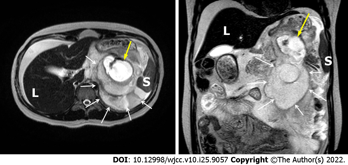
Figure 1 Magnetic resonance image showing a ruptured splenic artery (yellow arrow) with a huge hematoma (white arrow).
L: Liver; S: Spleen.
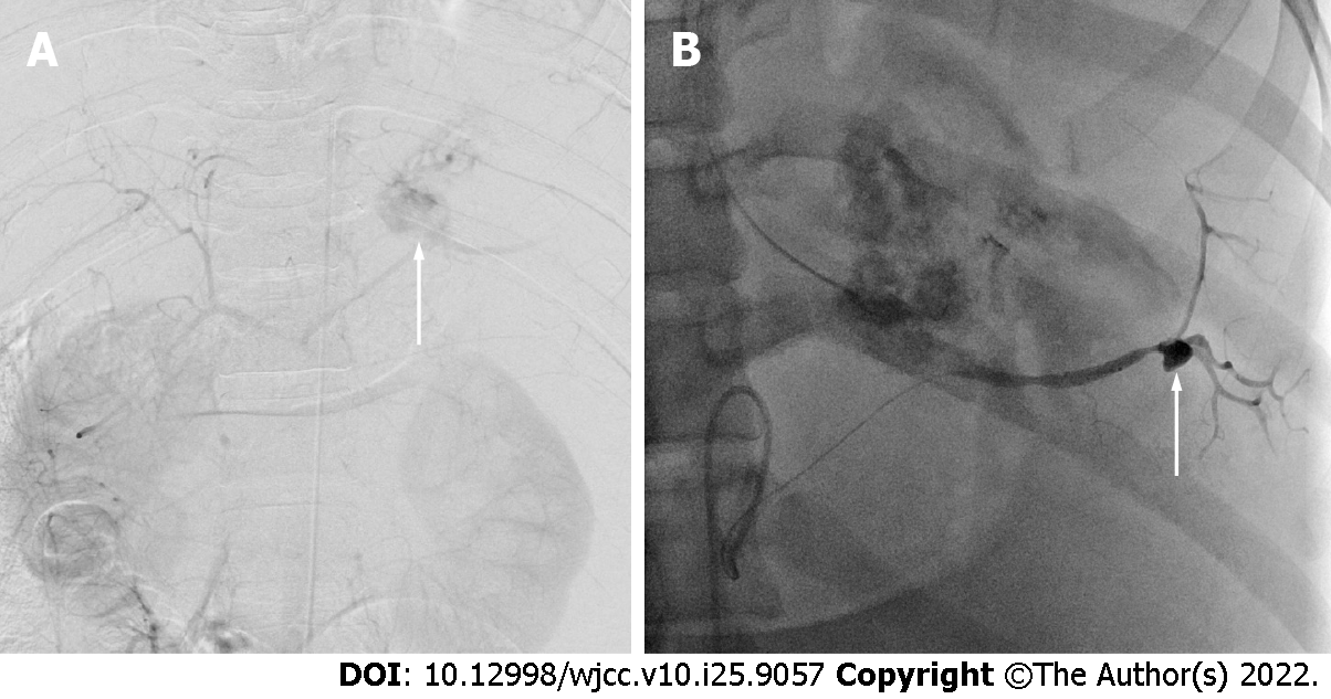
Figure 2 Digital arteriogram.
A: The image showing the abdominal aorta and the extravasation of contrast media from the mid-portion of the splenic artery (arrow); B: An additional aneurysm was found at the hilar portion of the splenic artery (arrow).
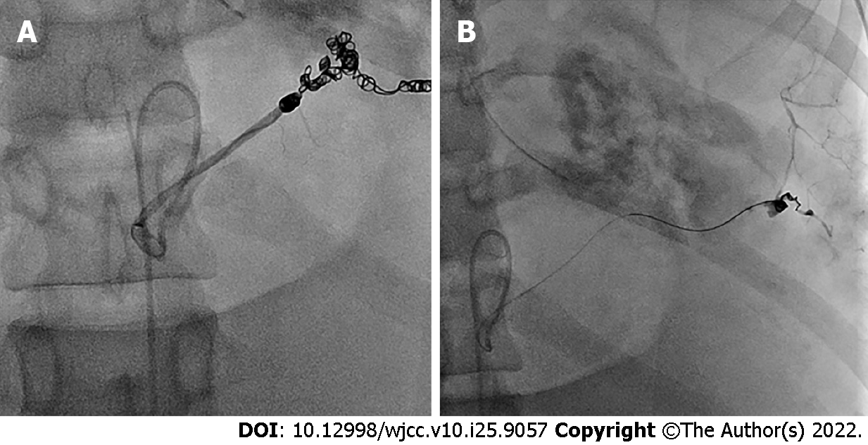
Figure 3 Postprocedural angiogram showing proximal embolization, the distal portion of a ruptured aneurysm (A), and another one at the hilar portion (B) by the micro-coil.
- Citation: Lee SH, Yang S, Park I, Im YC, Kim GY. Ruptured splenic artery aneurysms in pregnancy and usefulness of endovascular treatment in selective patients: A case report and review of literature. World J Clin Cases 2022; 10(25): 9057-9063
- URL: https://www.wjgnet.com/2307-8960/full/v10/i25/9057.htm
- DOI: https://dx.doi.org/10.12998/wjcc.v10.i25.9057


