Copyright
©The Author(s) 2022.
World J Clin Cases. Sep 6, 2022; 10(25): 8945-8953
Published online Sep 6, 2022. doi: 10.12998/wjcc.v10.i25.8945
Published online Sep 6, 2022. doi: 10.12998/wjcc.v10.i25.8945
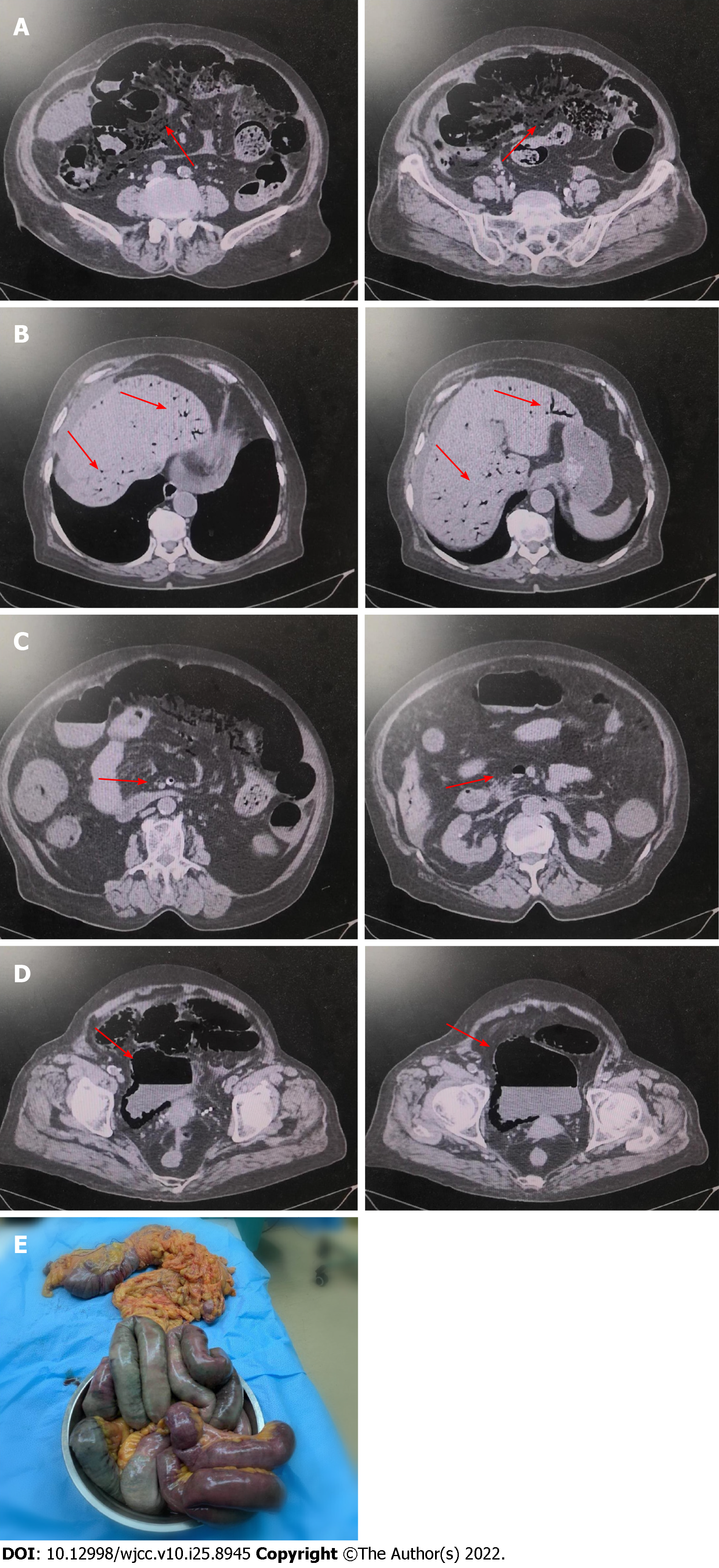
Figure 1 Computed tomography.
A: Pneumatosis intestinalis; B: Portal vein gas; C: Superior mesenteric artery and vein gas; D: Emphysematous cystitis; E: Necrotic intestinal canal and mesangial specimen resected.
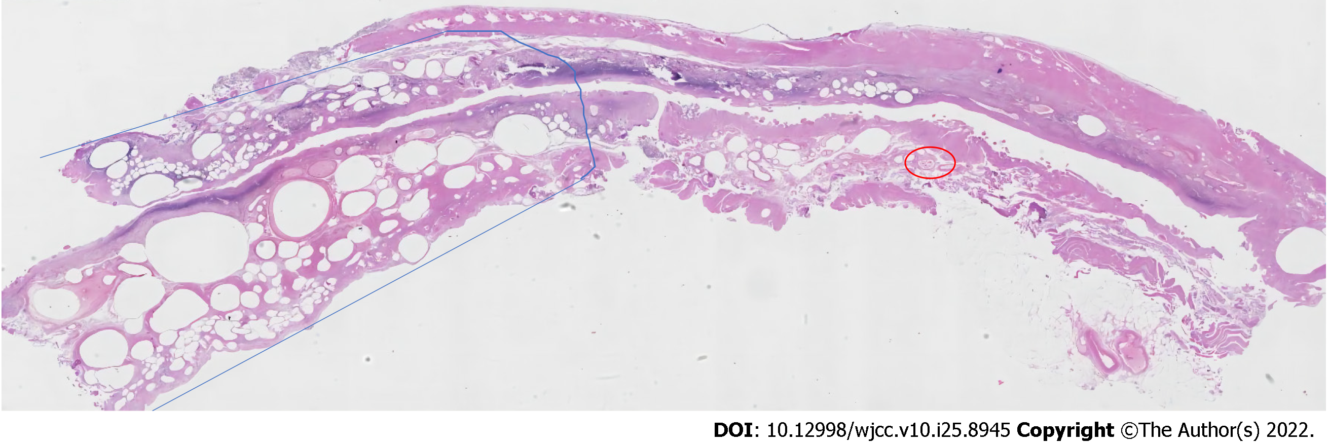
Figure 2 Histopathological findings: Muscular layer degenerating like honeycomb (blue curve area), thrombosis in blood vessels in the submucosa (red circle area) (HE, magnification × 4).
Figure 3 Computed tomography.
A: Portal vein gas disappeared after 7 d; B: Pneumatosis intestinalis disappeared after 7 d; C: Emphysematous cystitis after 7 d.
- Citation: Hu SF, Liu HB, Hao YY. Portal vein gas combined with pneumatosis intestinalis and emphysematous cystitis: A case report and literature review. World J Clin Cases 2022; 10(25): 8945-8953
- URL: https://www.wjgnet.com/2307-8960/full/v10/i25/8945.htm
- DOI: https://dx.doi.org/10.12998/wjcc.v10.i25.8945


