©The Author(s) 2022.
World J Clin Cases. Jan 14, 2022; 10(2): 733-740
Published online Jan 14, 2022. doi: 10.12998/wjcc.v10.i2.733
Published online Jan 14, 2022. doi: 10.12998/wjcc.v10.i2.733
Figure 1 Endoscopic findings (15 July 2020).
A: Multiple inflammation of the colon; B: Sigmoid colon ulcer with bleeding; C: Hemostasis under endoscopy.
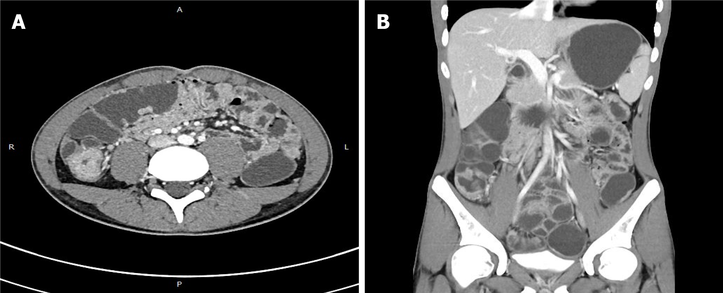
Figure 2 Computed tomography (18 July 2020).
A: Thickened walls of the small intestine; B: Thickened walls of colon.
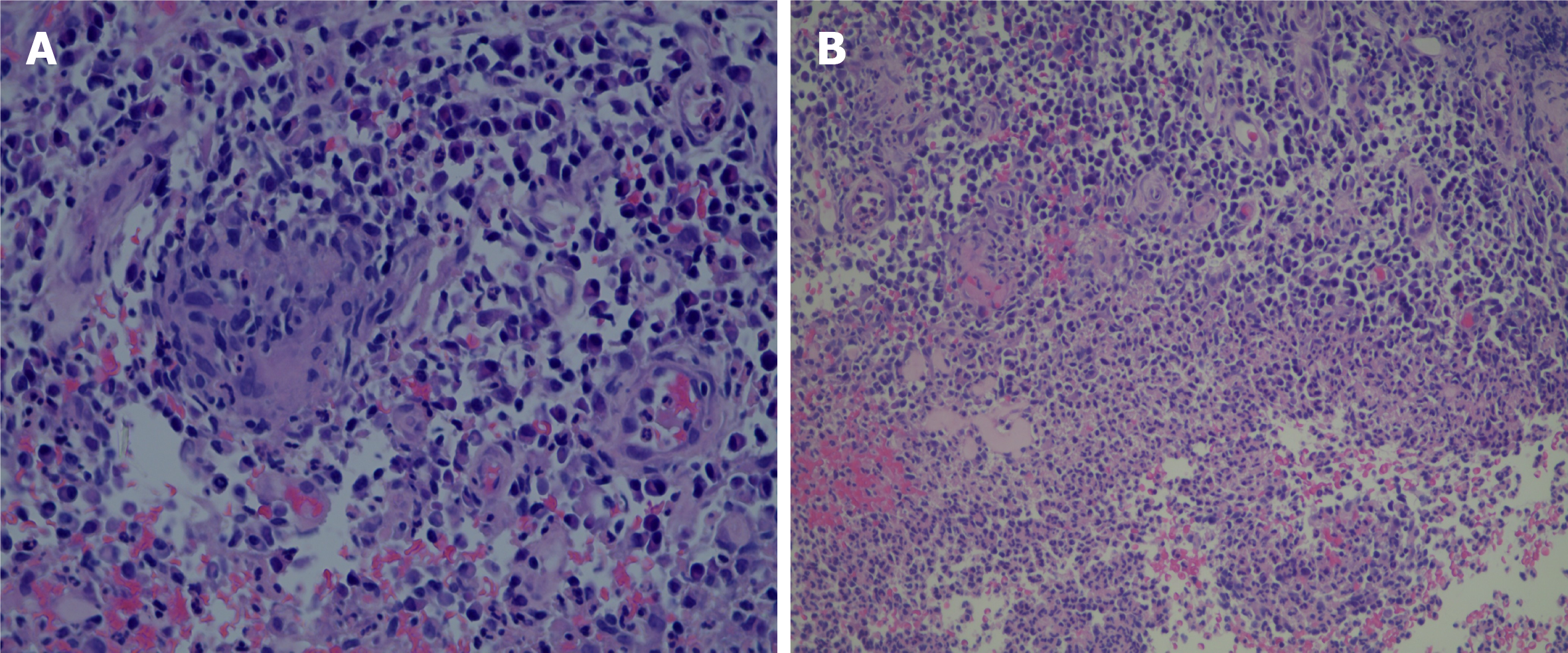
Figure 3 Pathology.
A: Acute on chronic inflammation with granulation tissue, consistent with Crohn's disease; B: Cytomegalovirus immunohistochemical staining and acid-fast staining were negative.
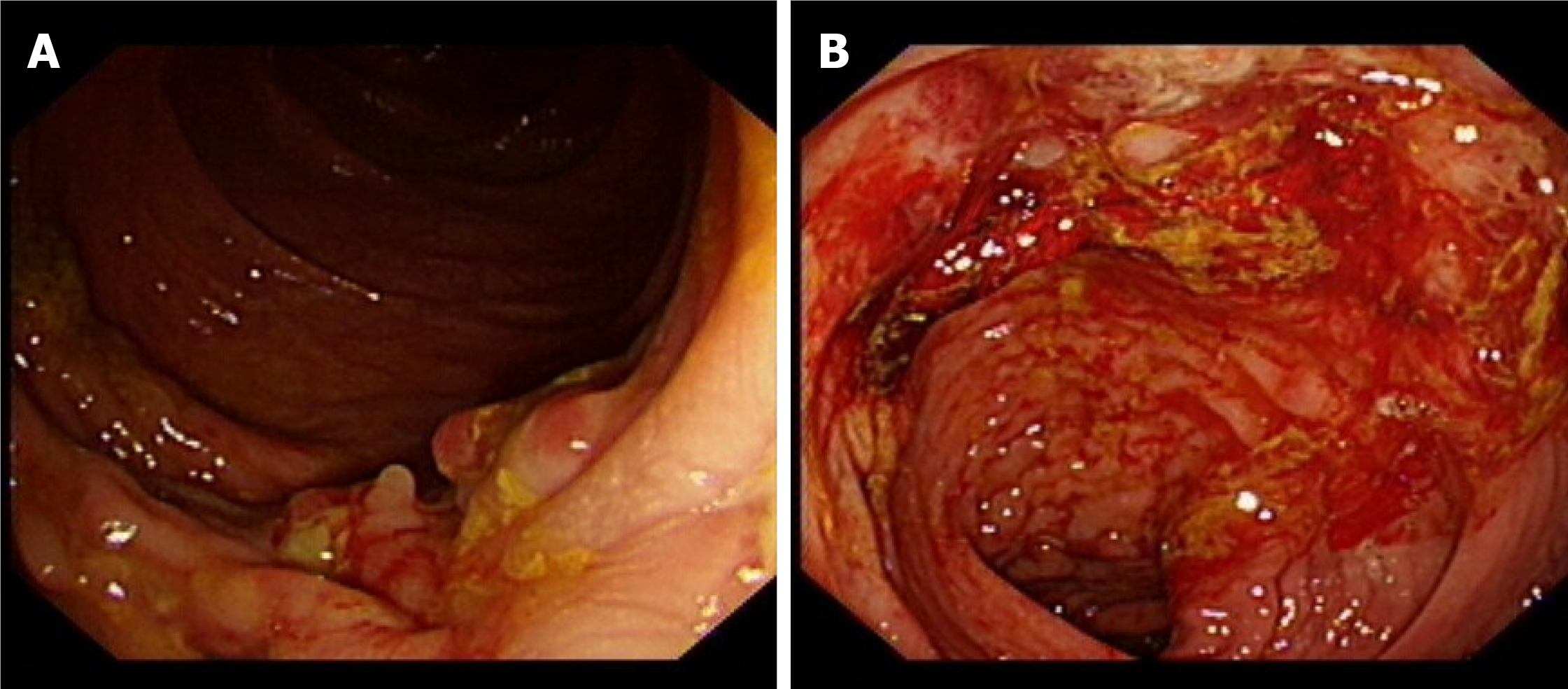
Figure 4 Endoscopic findings (25 July 2020).
A, B: Multiple ulcers with hemorrhage.
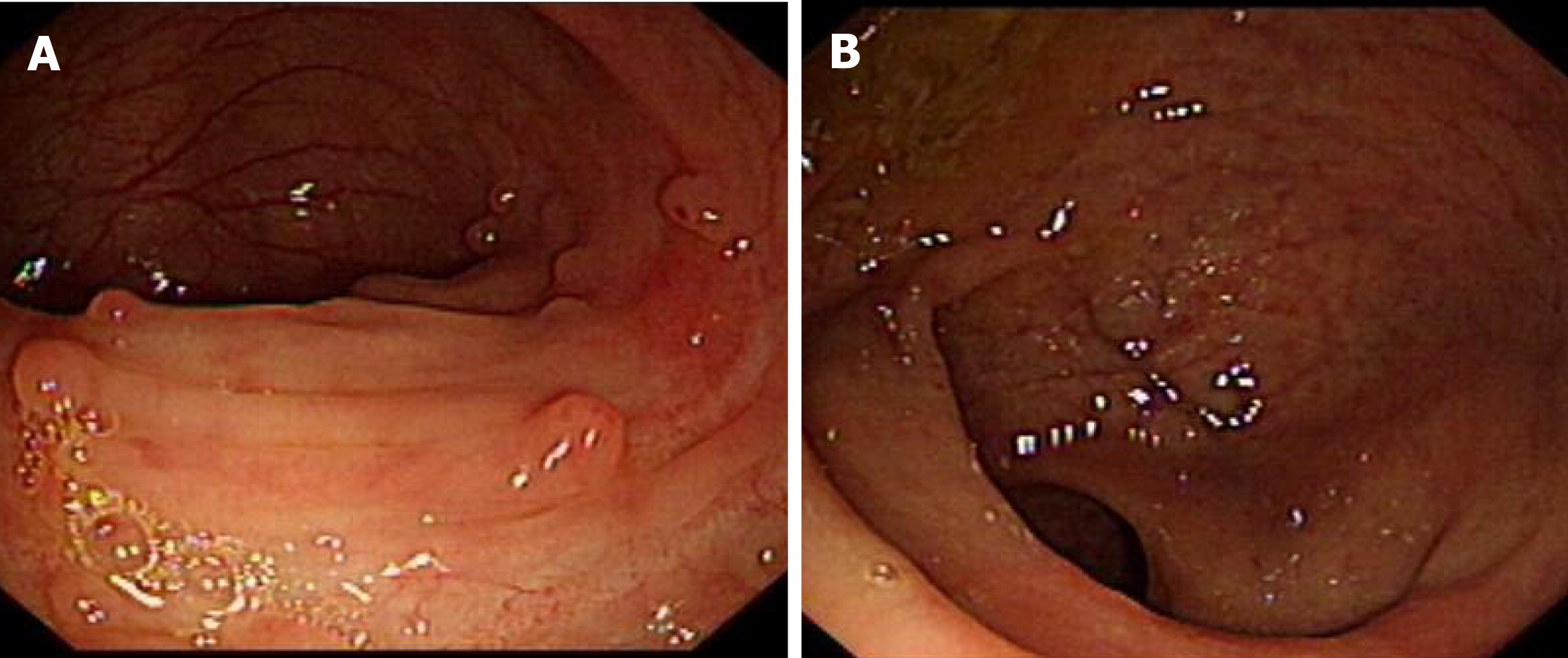
Figure 5 Endoscopic findings (8 wk after accelerated IFX induction).
A, B: Eight weeks after accelerated IFX induction therapy, colonoscopy showed mucosal healing. IFX: Anti-TNFα antibody.
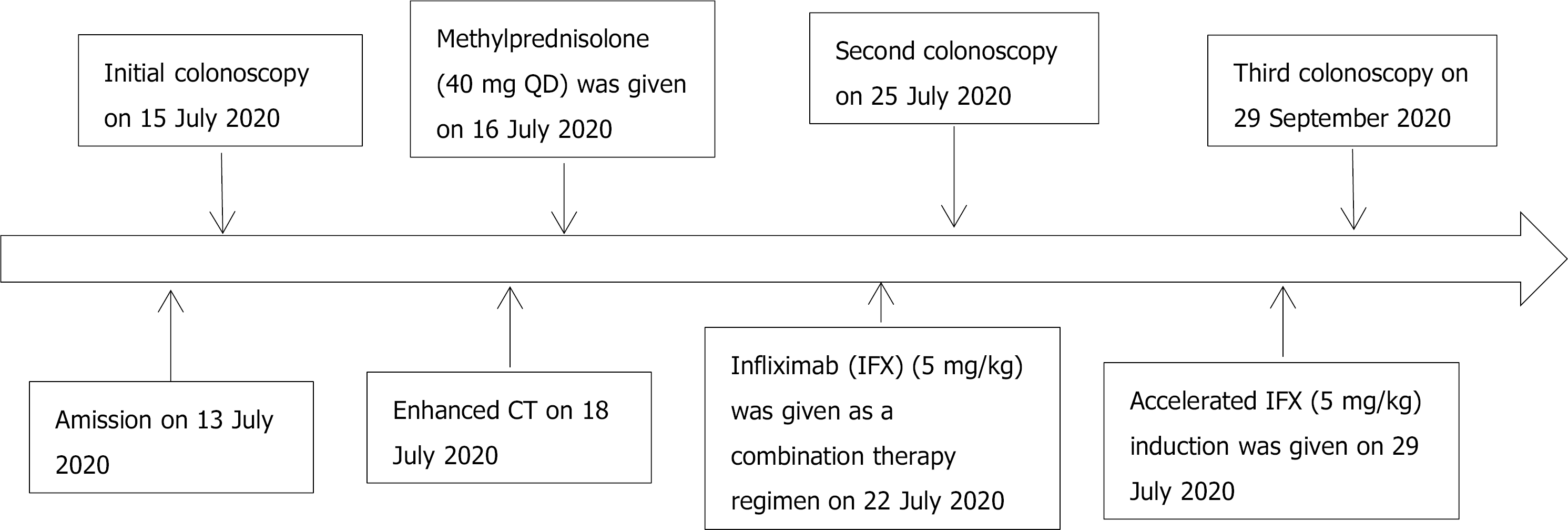
Figure 6 Timeline information in this case report.
CT: Computed tomography.
- Citation: Zeng J, Shen F, Fan JG, Ge WS. Accelerated Infliximab Induction for Severe Lower Gastrointestinal Bleeding in a Young Patient with Crohn’s Disease: A Case Report. World J Clin Cases 2022; 10(2): 733-740
- URL: https://www.wjgnet.com/2307-8960/full/v10/i2/733.htm
- DOI: https://dx.doi.org/10.12998/wjcc.v10.i2.733


