Copyright
©The Author(s) 2022.
World J Clin Cases. Jul 6, 2022; 10(19): 6672-6678
Published online Jul 6, 2022. doi: 10.12998/wjcc.v10.i19.6672
Published online Jul 6, 2022. doi: 10.12998/wjcc.v10.i19.6672
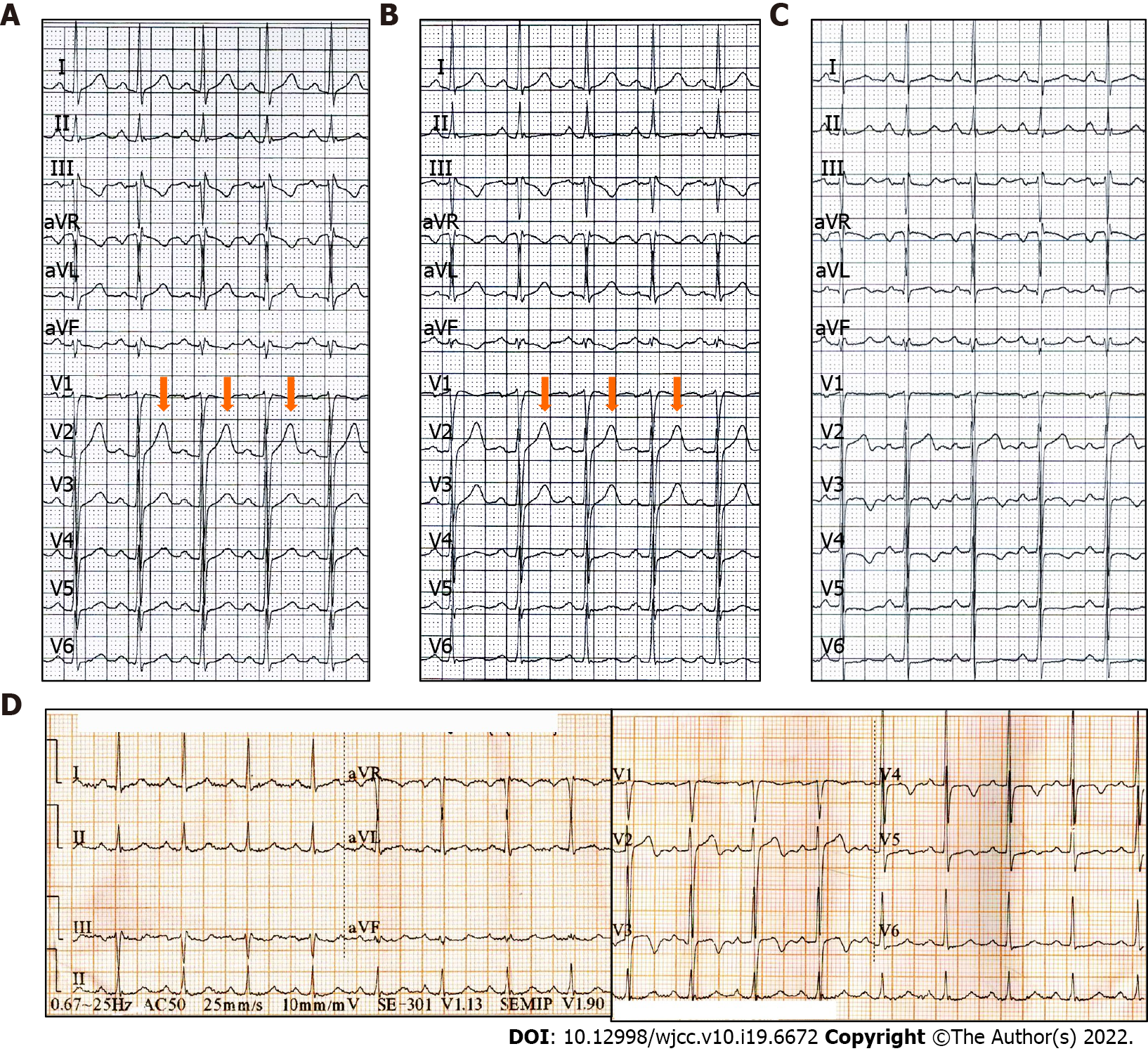
Figure 1 Preoperative electrocardiogram findings in a patient presenting with chest pain and exertional dyspnea.
A and B: The electrocardiograms (ECGs) are normal in the presence of angina; C: The ECG shows inverted or biphasic T-waves in the absence of angina; D: The ECG at admission in the absence of angina showing sinus tachycardia and T-wave changes.
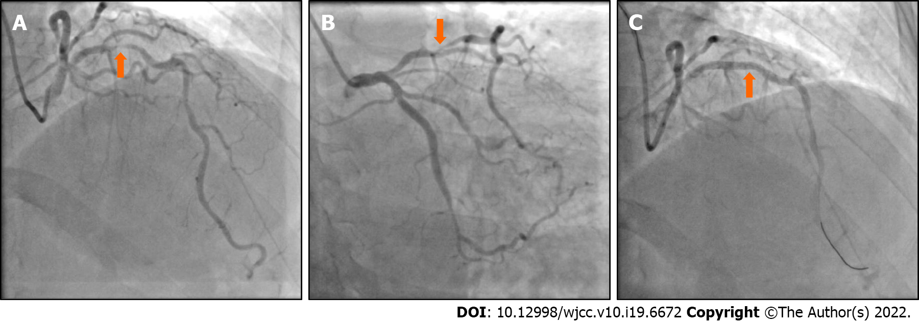
Figure 2 Coronary angiography findings in a patient presenting with chest pain and exertional dyspnea.
A and B: Coronary angiography (CAG) showing 90%-95% localized stenosis of the proximal left anterior descending (LAD) artery; C: CAG showing recovery of LAD flow after the percutaneous coronary intervention.
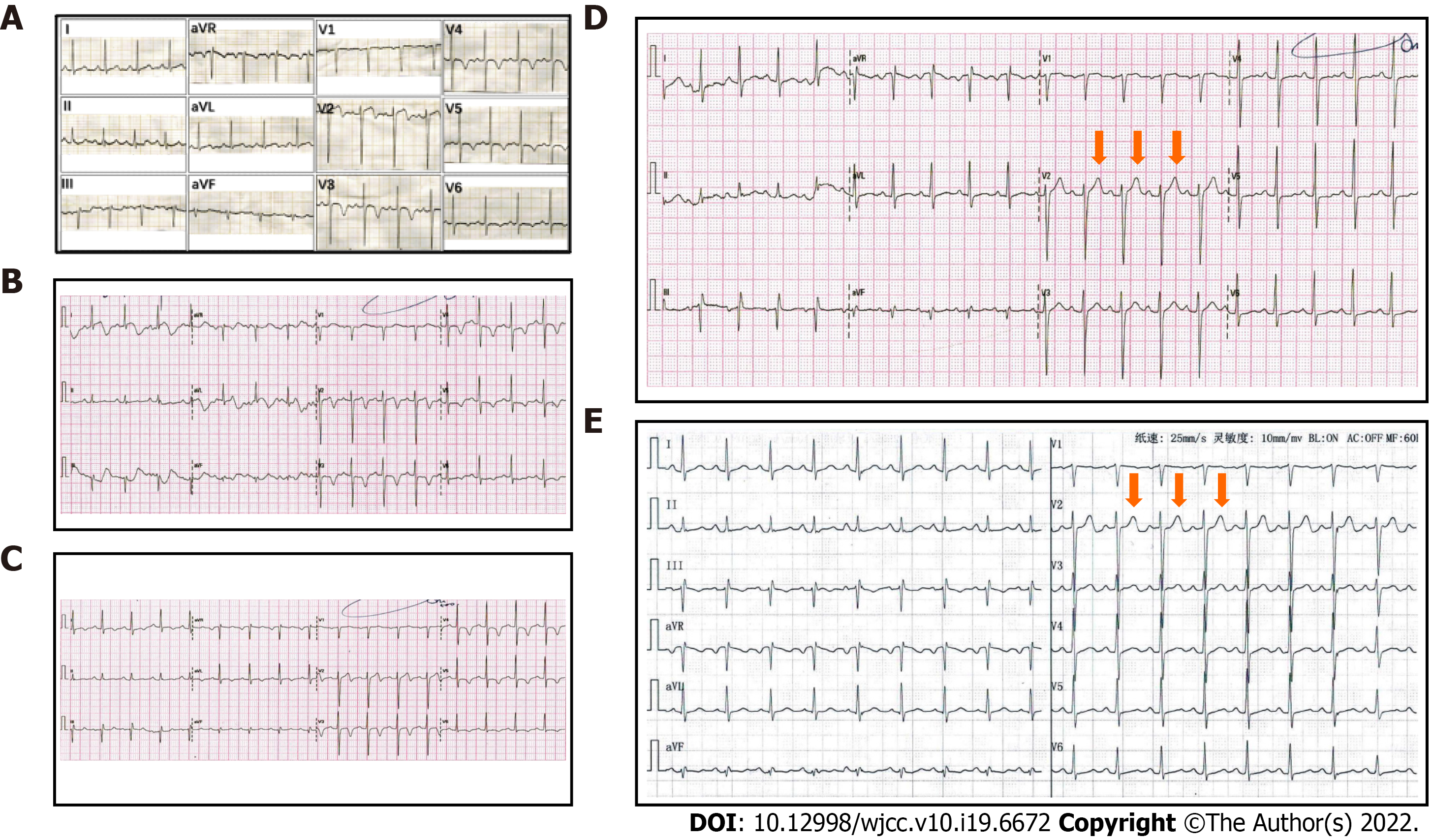
Figure 3 Electrocardiogram findings after percutaneous coronary intervention in a patient presenting with chest pain and exertional dyspnea.
A: Postoperative electrocardiograms (ECGs) showing inverted or biphasic T-waves at the extensive anterior precordial leads on the day of percutaneous coronary intervention (PCI); B: 1 d after PCI; C: 2 d after PCI; D: Postoperative ECGs showing normal T-waves on 3 d after PCI; E: 30 d after PCI.
- Citation: Tang N, Li YH, Kang L, Li R, Chu QM. Entire process of electrocardiogram recording of Wellens syndrome: A case report. World J Clin Cases 2022; 10(19): 6672-6678
- URL: https://www.wjgnet.com/2307-8960/full/v10/i19/6672.htm
- DOI: https://dx.doi.org/10.12998/wjcc.v10.i19.6672


