Copyright
©The Author(s) 2022.
World J Clin Cases. Jul 6, 2022; 10(19): 6520-6528
Published online Jul 6, 2022. doi: 10.12998/wjcc.v10.i19.6520
Published online Jul 6, 2022. doi: 10.12998/wjcc.v10.i19.6520
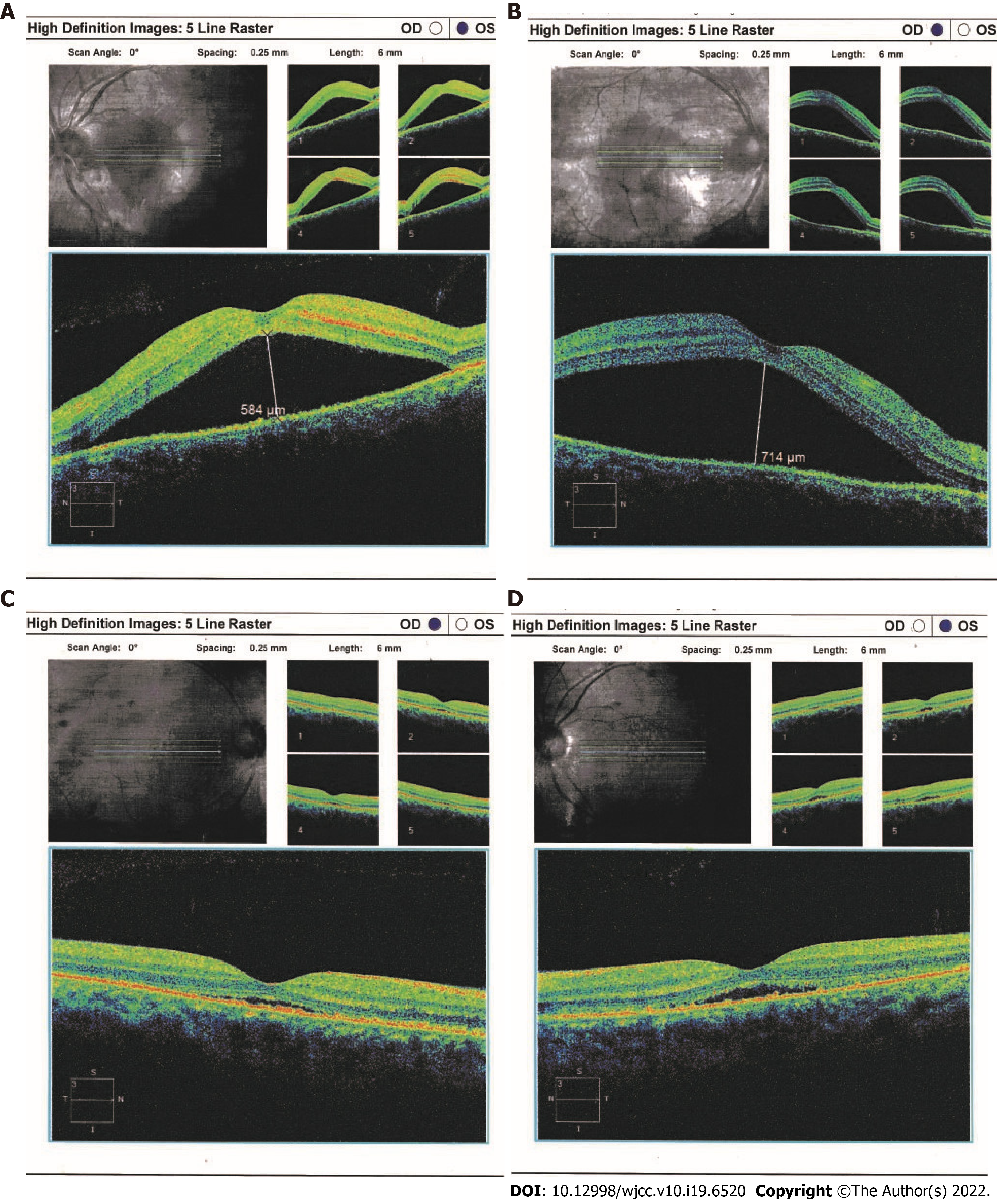
Figure 1 Ocular coherence tomography and ophthalmoscopy of both eyes.
Serous retinal detachment (arrows) were obvious by ocular coherence tomography in the right (A) and left (B) eyes at initial presentation (May 22, 2018); C and D: After treatment with a systemic corticosteroid and an immunosuppressive agent, the detachment was improved in both eyes (June 19, 2018).
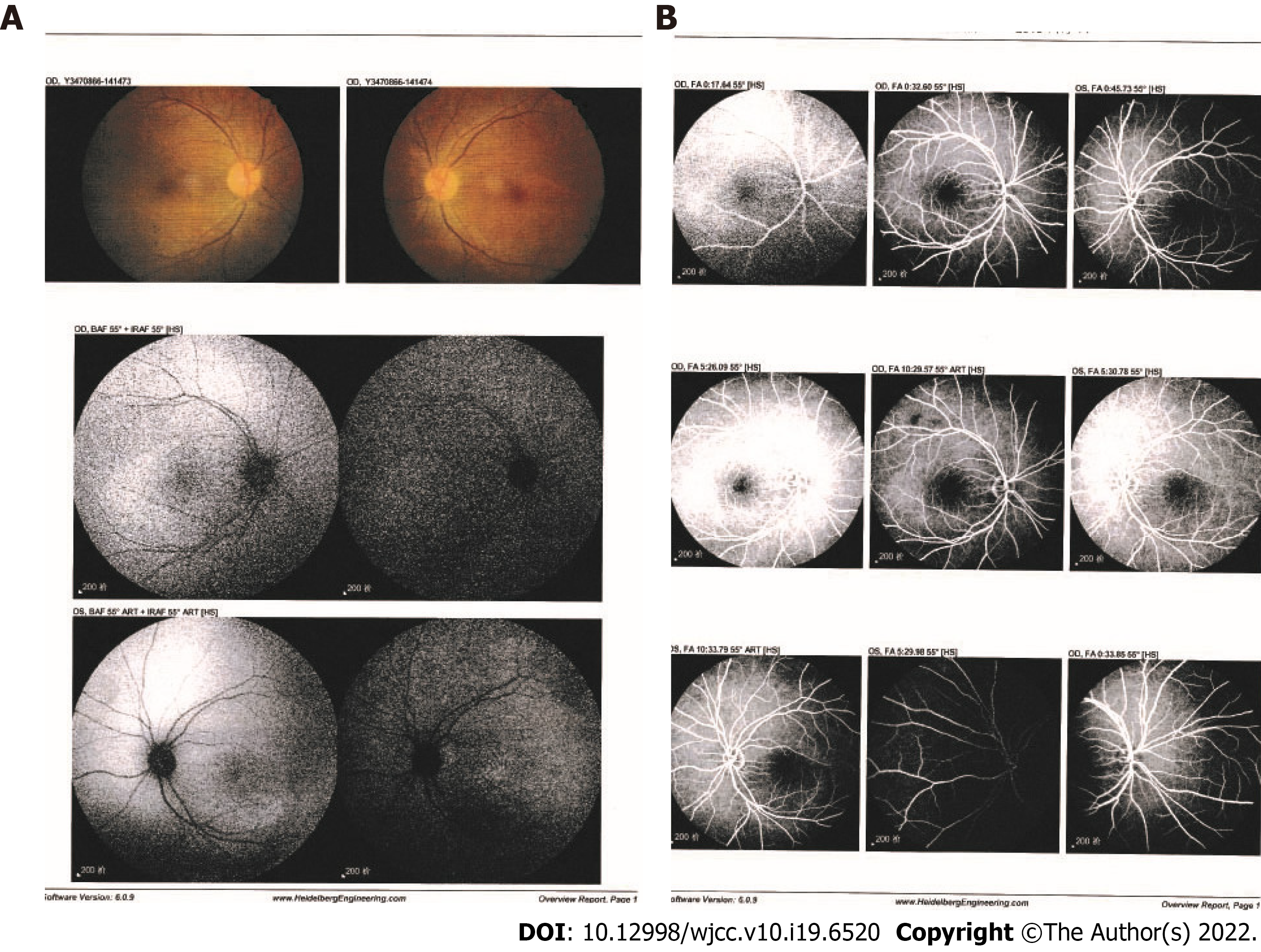
Figure 2 Fundus fluorescein angiography and indocyanine green angiography findings.
Indocyanine green angiography (A) and fluorescein angiography (B) images of the patient after hospitalization showed no leakage from choroidal vessels (June 14, 2018).
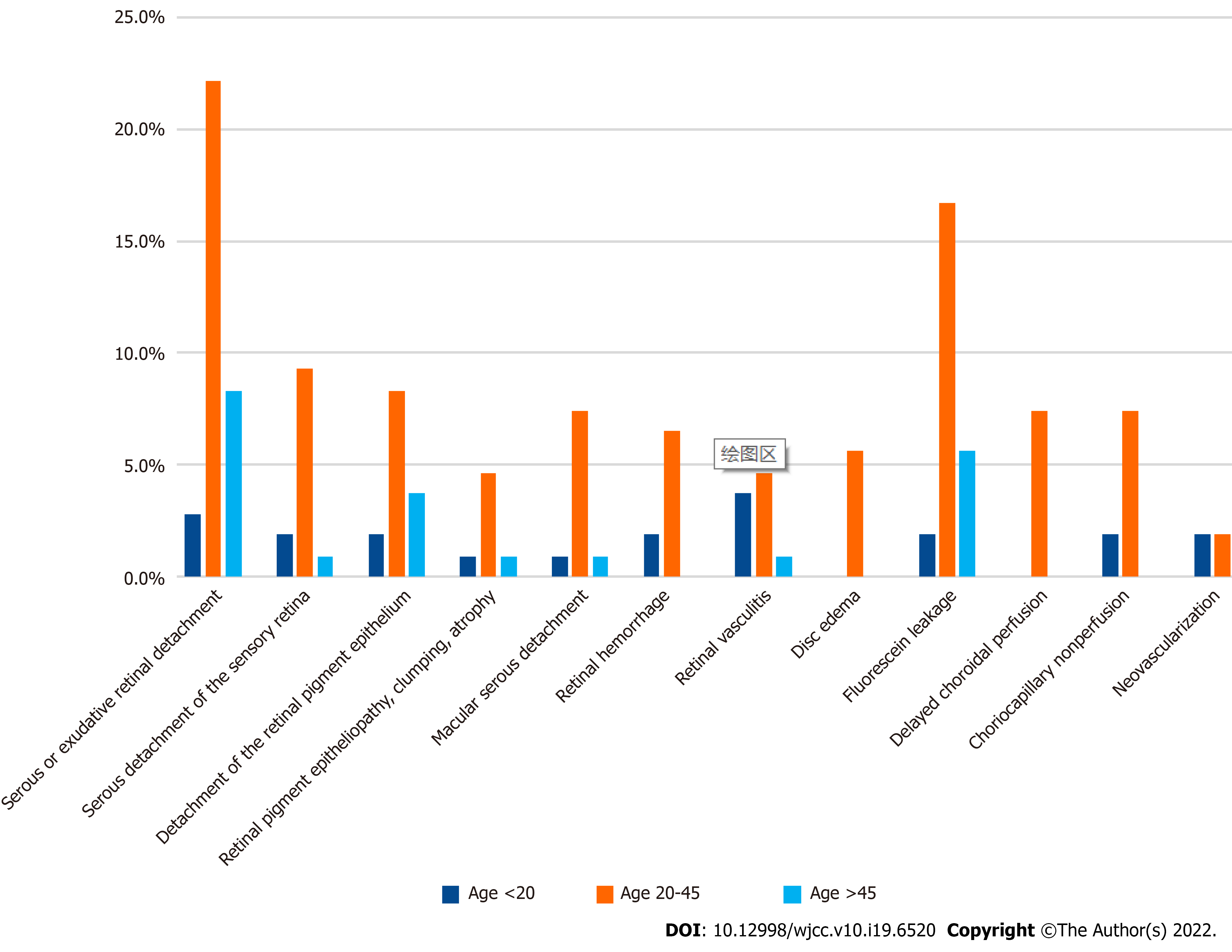
Figure 3 Frequencies of ocular imaging features of lupus choroidopathy in literature review.
The patients were grouped by age at ocular involvement.
- Citation: Yao Y, Wang HX, Liu LW, Ding YL, Sheng JE, Deng XH, Liu B. Acute choroidal involvement in lupus nephritis: A case report and review of literature. World J Clin Cases 2022; 10(19): 6520-6528
- URL: https://www.wjgnet.com/2307-8960/full/v10/i19/6520.htm
- DOI: https://dx.doi.org/10.12998/wjcc.v10.i19.6520


