Copyright
©The Author(s) 2022.
World J Clin Cases. Jun 26, 2022; 10(18): 6241-6246
Published online Jun 26, 2022. doi: 10.12998/wjcc.v10.i18.6241
Published online Jun 26, 2022. doi: 10.12998/wjcc.v10.i18.6241
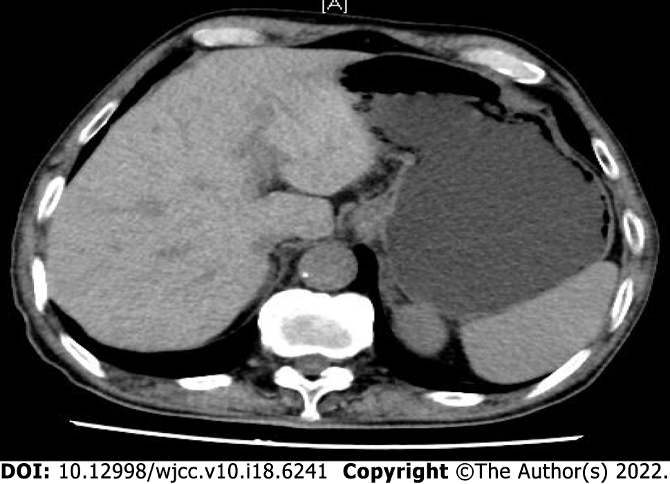
Figure 1 Full abdominal enhanced computed tomography.
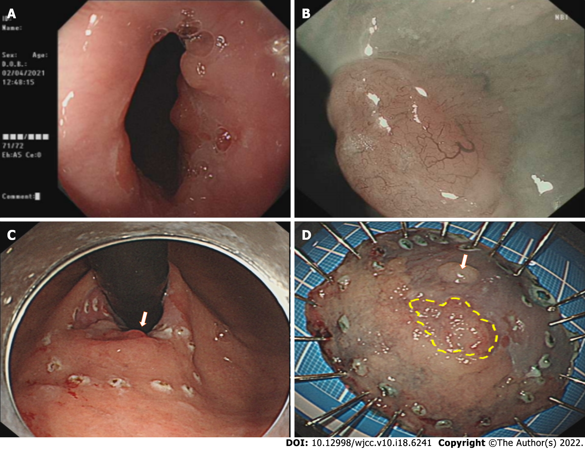
Figure 2 Endoscopic submucosal dissection.
A: Cardia mass; B: Submucosal injection; C: Tick to mark the lesion area; D: Lesion specimen.
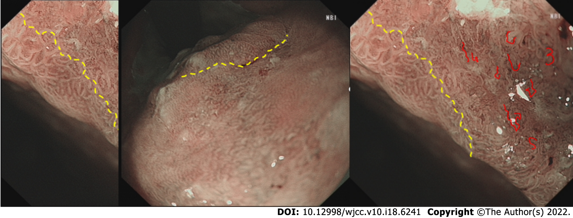
Figure 3 Narrow-band imaging.
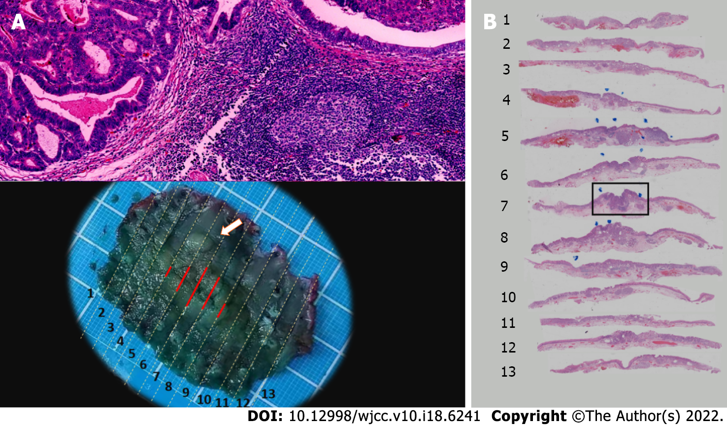
Figure 4 Pathology of cardia endoscopic submucosal dissection specimens.
A: Moderately differentiated adenocarcinoma of the cardia; B: Esophageal neuroendocrine tumors (G3).
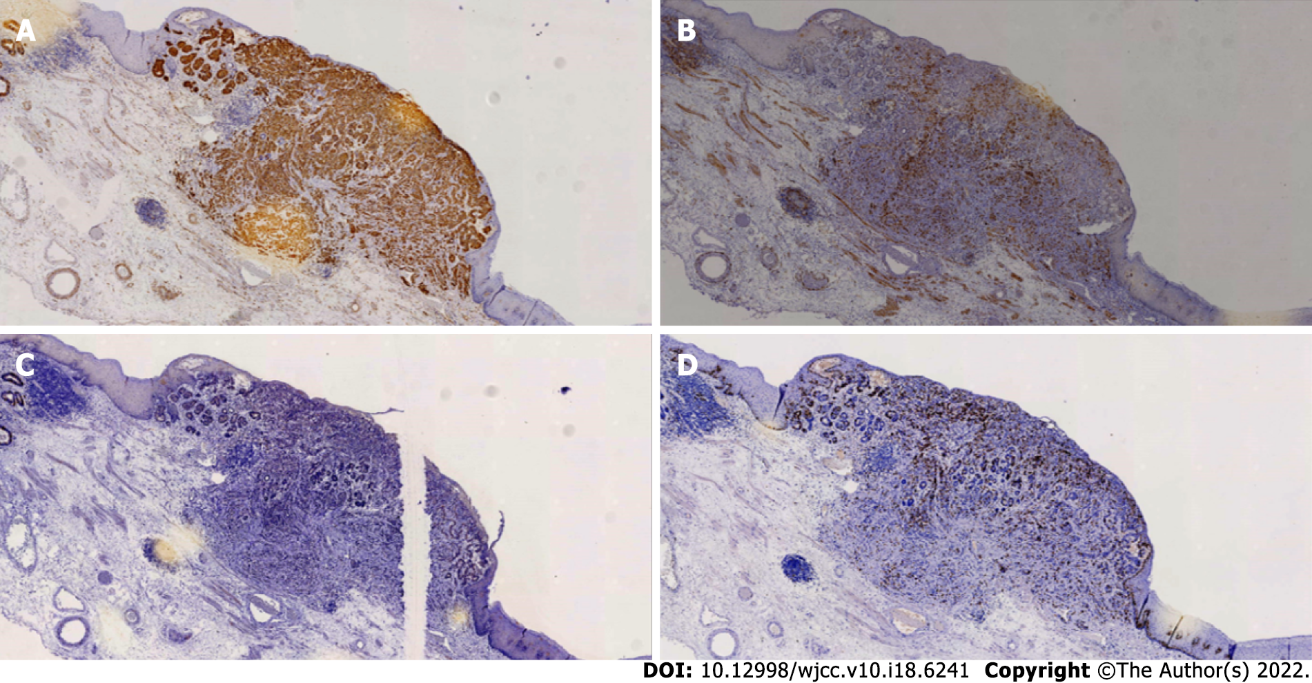
Figure 5 Immunohistochemical staining of esophageal neuroendocrine tumors.
A: Syn; B: CD56; C: CgA; D: Ki-67 (25%+).
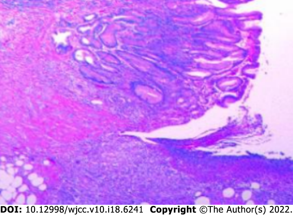
Figure 6 Representative pathology of excised gastric specimens.
- Citation: Kong ZZ, Zhang L. Esophagogastric junctional neuroendocrine tumor with adenocarcinoma: A case report. World J Clin Cases 2022; 10(18): 6241-6246
- URL: https://www.wjgnet.com/2307-8960/full/v10/i18/6241.htm
- DOI: https://dx.doi.org/10.12998/wjcc.v10.i18.6241


