Copyright
©The Author(s) 2022.
World J Clin Cases. Jun 16, 2022; 10(17): 5910-5915
Published online Jun 16, 2022. doi: 10.12998/wjcc.v10.i17.5910
Published online Jun 16, 2022. doi: 10.12998/wjcc.v10.i17.5910
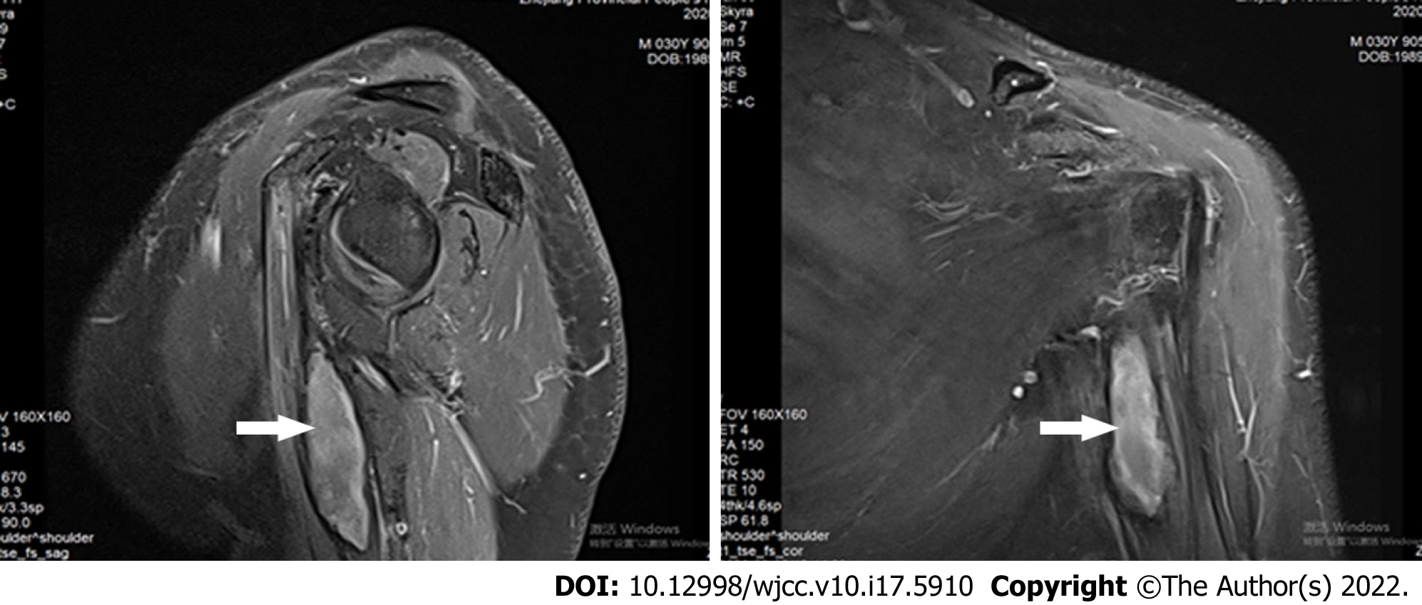
Figure 1 Magnetic resonance images of the left upper limb.
Sagittal and coronal views of T2-weighted images showed a focal mass on left coracobrachialis muscle (white arrow).
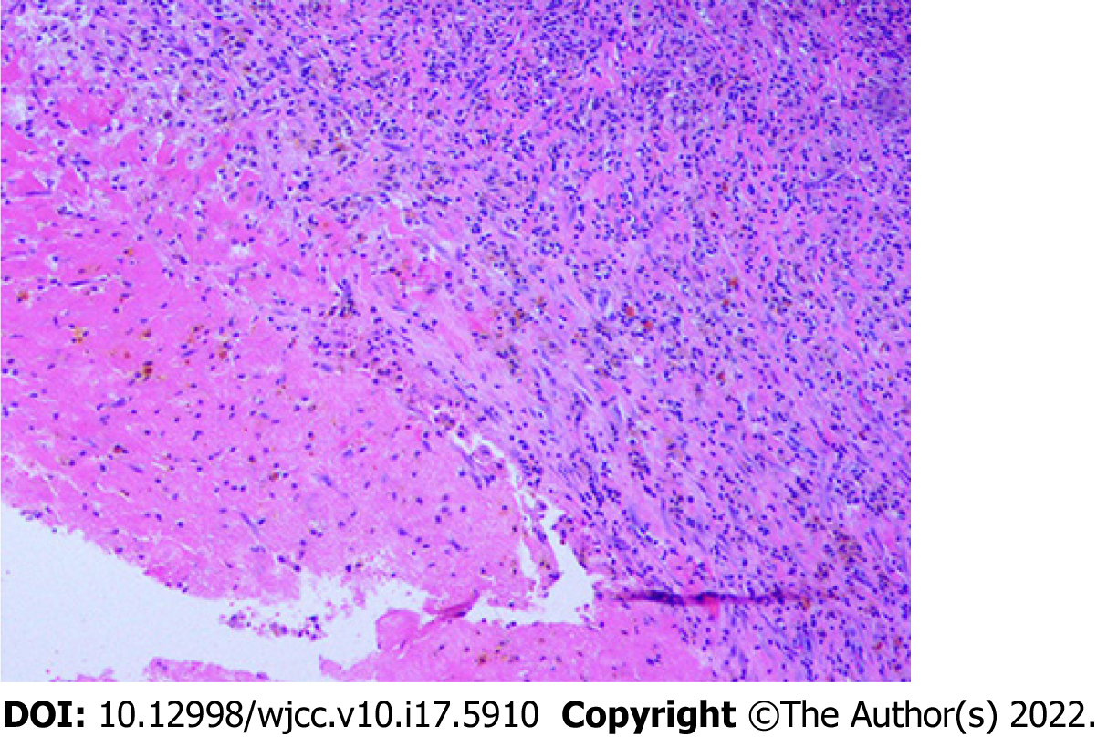
Figure 2
Histopathological examination of the resected mass showed inflammatory infiltration with hemosiderin pigments and fibrosis (hematoxylin and eosin stain, × 100).
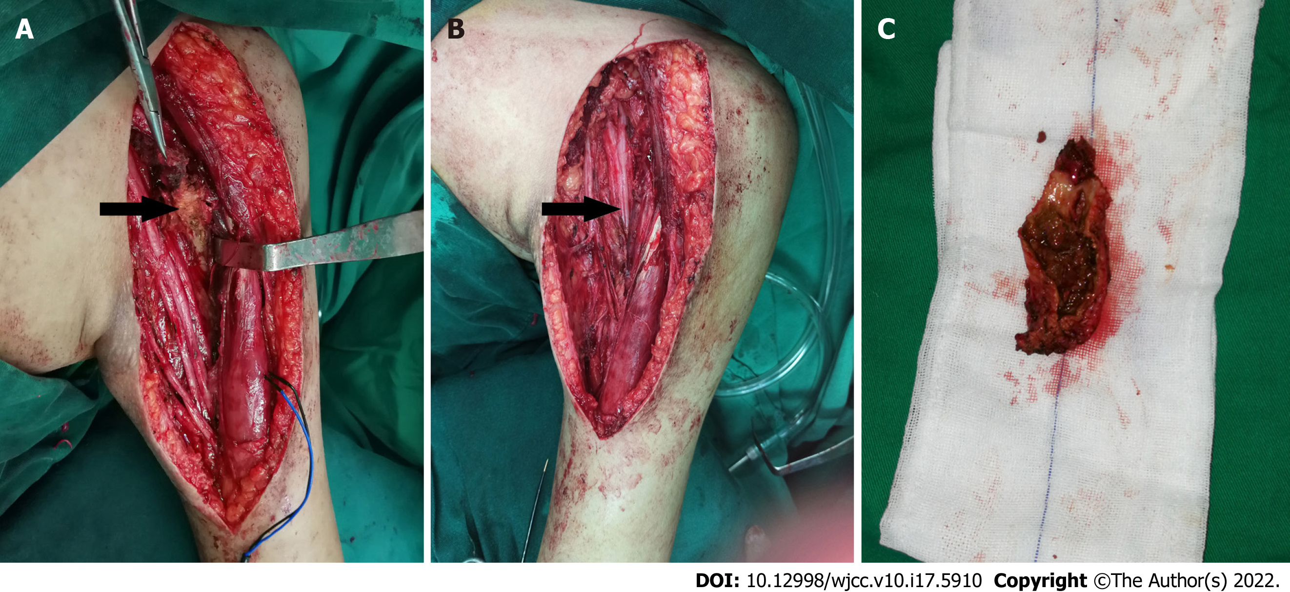
Figure 3 Intraoperative findings of the patient.
A: An organized mass was exposed prior to excision (black arrow); B: Brachial plexus neurolysis was performed after the mass resection; C: The cut surface of the resected specimen indicated an organized chronic hematoma rather than a neoplasm.
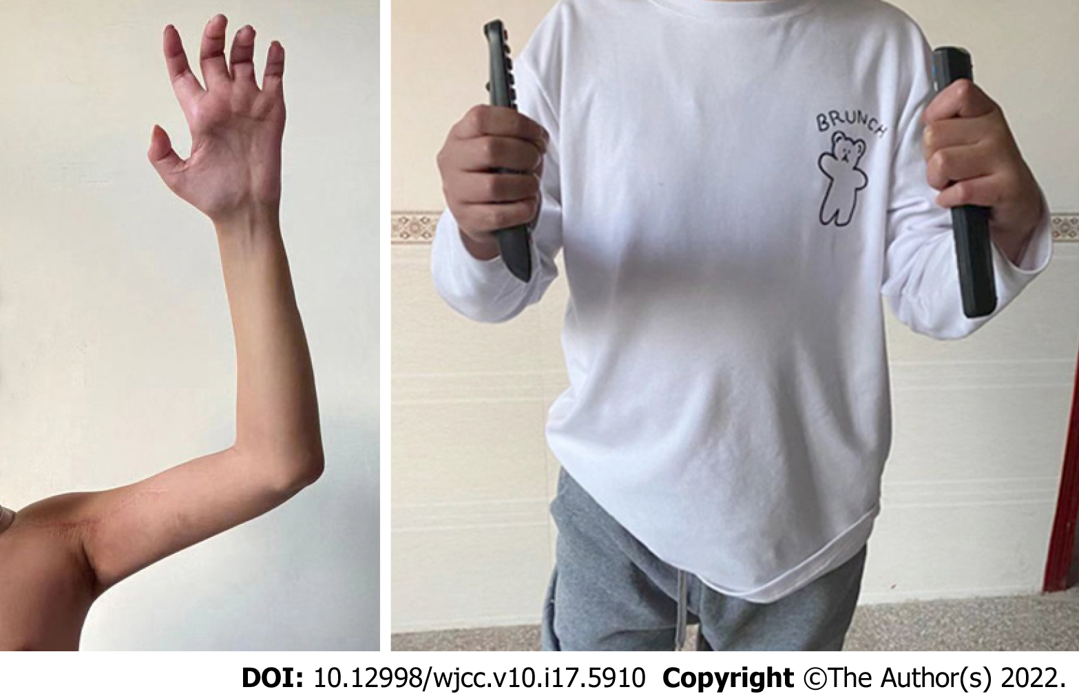
Figure 4
A 12 mo follow-up assessment demonstrated definite improvement in the left shoulder abduction, elbow flexion and grip strength after successful surgery.
- Citation: Guo EQ, Yang XD, Lu HR. Tumor-like disorder of the brachial plexus region in a patient with hemophilia: A case report. World J Clin Cases 2022; 10(17): 5910-5915
- URL: https://www.wjgnet.com/2307-8960/full/v10/i17/5910.htm
- DOI: https://dx.doi.org/10.12998/wjcc.v10.i17.5910


