©The Author(s) 2022.
World J Clin Cases. Jun 16, 2022; 10(17): 5748-5755
Published online Jun 16, 2022. doi: 10.12998/wjcc.v10.i17.5748
Published online Jun 16, 2022. doi: 10.12998/wjcc.v10.i17.5748
Figure 1 Preoperative computed tomography of the cervical spine shows lesions of the C7 vertebral body and C7/T1 intervertebral disc.
A: Three-dimensional computed tomography (CT) sagittal C7 vertebral lesions; B: Three-dimensional CT coronal C7 vertebral lesions; C: Three-dimensional CT axial C7 vertebral lesions.
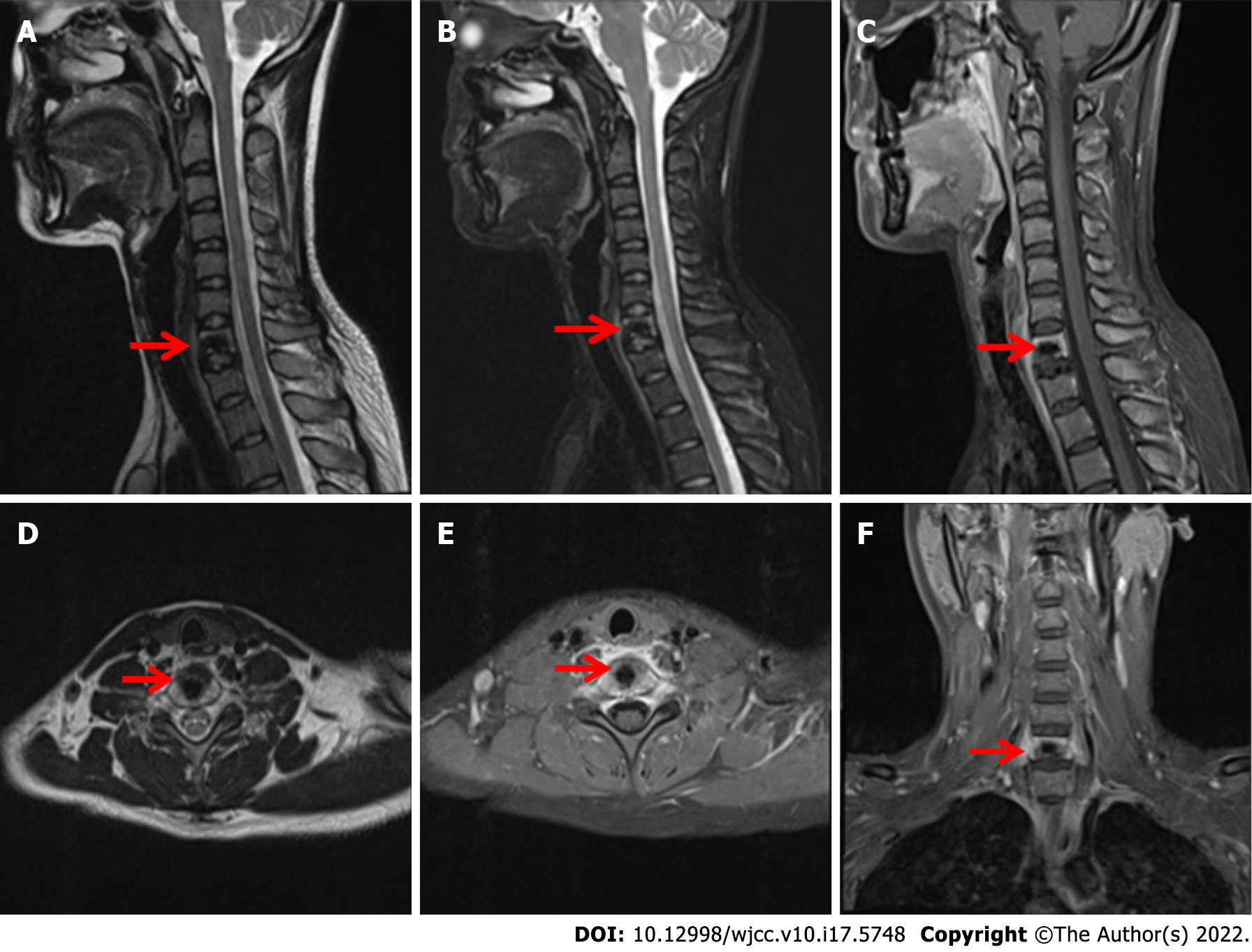
Figure 2 Magnetic resonance imaging of the cervical vertebra shows a patchy, abnormal signal shadow of the C7 vertebral body, swelling of adjacent soft tissue, uneven enhancement after enhancement, and no abnormal signal of the cervical spinal cord.
A, D: T1-weighted images; B: T2-weighted images; C, E and F: Fat suppression images.
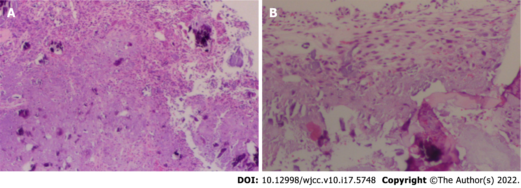
Figure 3 Visible under the microscope: Medullary bone and bone trabeculae, part of the nucleus pulposus and abundant bone marrow were observed.
Cartilaginous myxoid stroma, multifocal proliferating fusiform fibrous tissue with considerable calcium deposition and multinucleated giant cells are on the side. A, B: Hematoxylin and eosin (HE) staining, original magnification × 10.
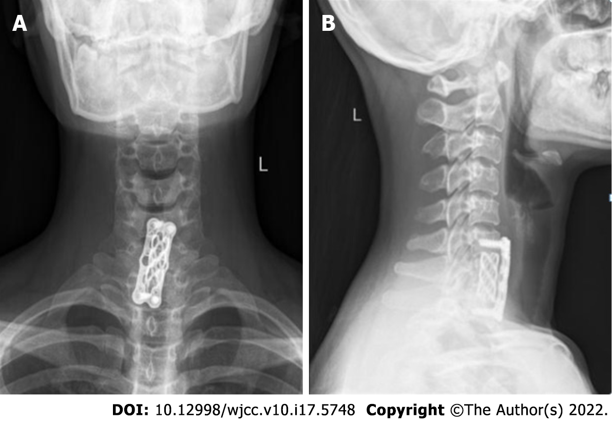
Figure 4 Postoperative X-ray of cervical vertebra.
A, B: Nine months after the surgery, the cervical spine was re-examined in the anteroposterior and lateral positions and showed that the internal fixation position was good.
- Citation: Li C, Li S, Hu W. Chondromyxoid fibroma of the cervical spine: A case report. World J Clin Cases 2022; 10(17): 5748-5755
- URL: https://www.wjgnet.com/2307-8960/full/v10/i17/5748.htm
- DOI: https://dx.doi.org/10.12998/wjcc.v10.i17.5748


