Copyright
©The Author(s) 2022.
World J Clin Cases. Jun 16, 2022; 10(17): 5702-5707
Published online Jun 16, 2022. doi: 10.12998/wjcc.v10.i17.5702
Published online Jun 16, 2022. doi: 10.12998/wjcc.v10.i17.5702
Figure 1 Expression of GPI-anchored proteins in patient peripheral blood cells before and after allo-HSCT.
A: Before transplantation: expression of (left) CD59 on red cells, (center) CD24/FLAER on neutrophils, and (right) CD14/FLAER on monocytes. There is a mosaic of cells with normal expression of GPI-anchored proteins and cells with reduced (type II) or completely lacking expression (type III) of GPI-anchored proteins; B: After transplantation, CD59 was expressed on 99.8% of red cells (left), CD24/FLAER on 100% of neutrophils (center) and CD14/FLAER on 100% of monocytes (right).
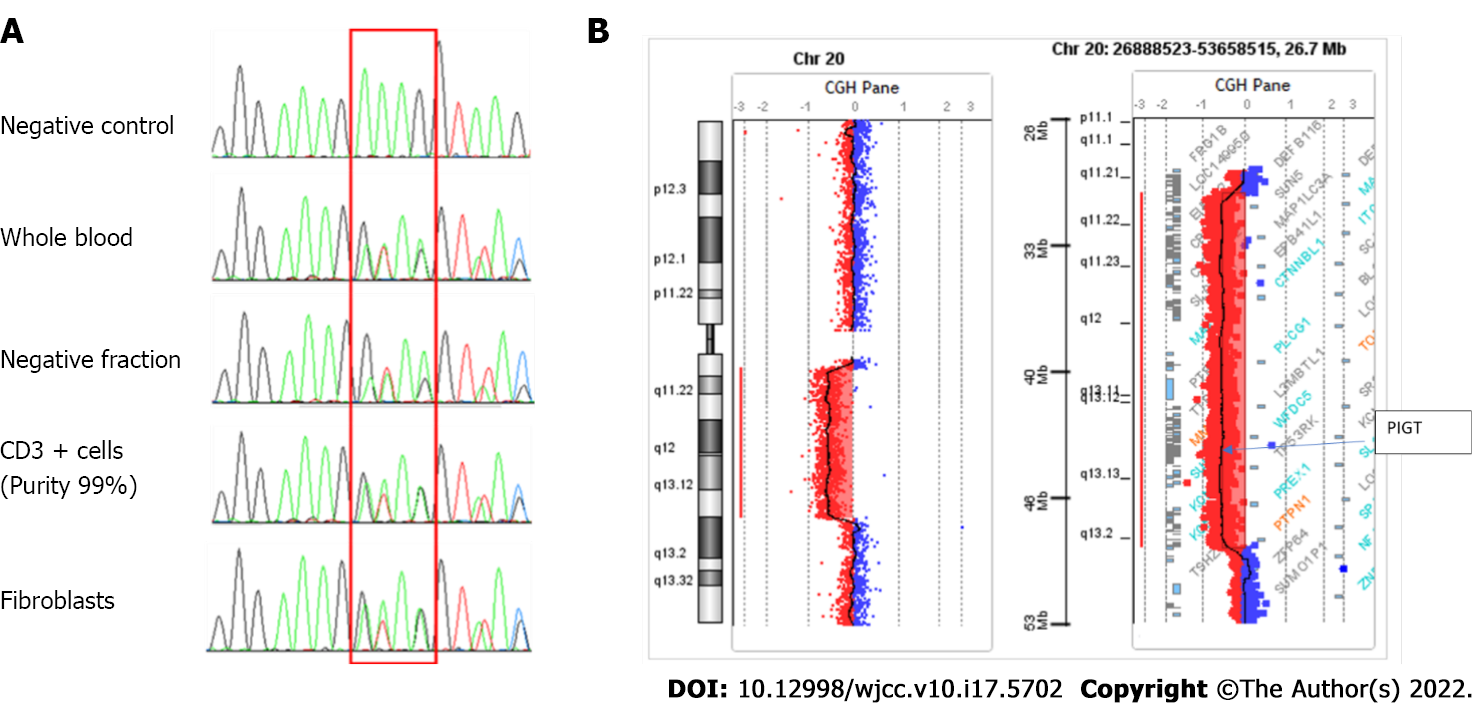
Figure 2 Genetic analysis.
A: Genetic sequencing. Cell sorting was performed on blood samples (Robosep, Easy sep CD3 whole blood positive selection kit®). The positive fraction consisted of a T lymphocyte population (purity: 99%), and the negative fraction included B lymphocytes, natural killers, monocytes and polymorphonuclear cells. DNA was extracted from these two fractions using a Qiagen DNA minikit®. The entire PIGT gene was sequenced by the Sanger method (Big Dye Terminator v3.1, Life Technologies) using primers for each exon; B: CGH array (Agilent, Sure Print G3 Human CGH Microarray 4x180K) performed on DNA extracted from bone marrow samples highlights a large somatic deletion of 18 Mb from 20q11.21 to 20q13.13, removing the entire PIGT gene.
- Citation: Schenone L, Notarantonio AB, Latger-Cannard V, Fremeaux-Bacchi V, De Carvalho-Bittencourt M, Rubio MT, Muller M, D'Aveni M. Allogeneic stem cell transplantation-A curative treatment for paroxysmal nocturnal hemoglobinuria with PIGT mutation: A case report. World J Clin Cases 2022; 10(17): 5702-5707
- URL: https://www.wjgnet.com/2307-8960/full/v10/i17/5702.htm
- DOI: https://dx.doi.org/10.12998/wjcc.v10.i17.5702


