©The Author(s) 2022.
World J Clin Cases. May 26, 2022; 10(15): 5057-5063
Published online May 26, 2022. doi: 10.12998/wjcc.v10.i15.5057
Published online May 26, 2022. doi: 10.12998/wjcc.v10.i15.5057
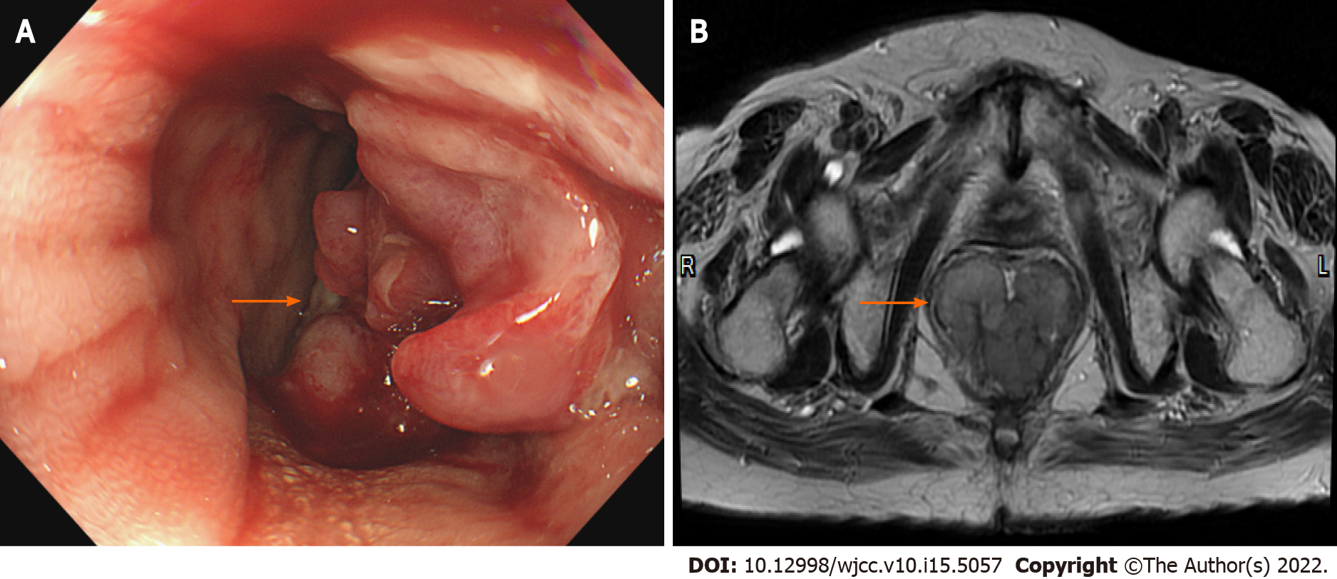
Figure 1 Initial colonoscopy and magnetic resonance imaging.
A: Colonoscopy showed a polypoid mass about 6 cm in size at rectum; B: Magnetic resonance imaging showed a lobulated mixed-signal intensity mass at the rectum (5.5 cm × 4.8 cm in size, 3.5 cm above the anal verge) on the T2-weighted axial image.
Figure 2 Histology image of the biopsied tissues.
A: Neoplastic cells showed epithelioid morphology (hematoxylin-eosin staining, 400 ×); B: Neoplastic cells showed focally positive staining for HMB-45 (immunohistochemistry staining, 40 ×); C: Neoplastic cells showed positive staining for Melan-A (immunohistochemistry staining, 40 ×); D: Neoplastic cells showed positive staining for S-100 (immunohistochemistry staining, 40 ×).
Figure 3 Computed tomography and positron emission tomography/computed tomography for pelvic imaging in anorectal melanoma.
A and B: The (A) axial computed tomography (CT) and (B) positron emission tomography/CT (PET/CT) showed Intense FDG-avidity over the rectum, suspicious for malignancy; C and D: The (C) axial CT and (D) PET/CT showed one mild FDG-avidity over the pre-sacral space, suspicious nodal metastasis.
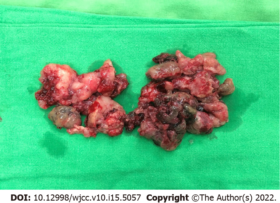
Figure 4 The first wide local excision was performed with snares, to cut off parts of the polypoid mass piece by piece until the protruding mass is resected.
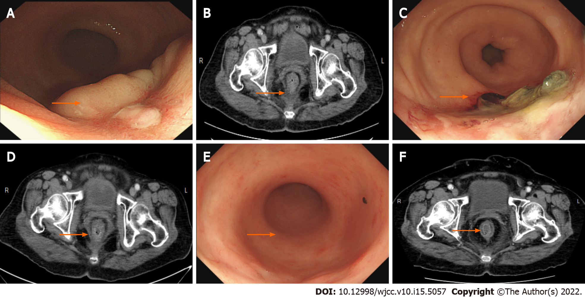
Figure 5 Following-up colonoscopy and computed tomography.
A and B: The (A) colonoscopy and (B) computed tomography (CT) showed 1 mo after radiotherapy [before the second wide local excision (WLE)]; C and D: The (C) colonoscopy and (D) CT showed 3 mo after the second WLE; E and F: The (E) colonoscopy and (F) CT showed 2 yr after the second WLE.
- Citation: Chiu HT, Pu TW, Yen H, Liu T, Wen CC. Is repeat wide excision plus radiotherapy of localized rectal melanoma another choice before abdominoperineal resection? A case report. World J Clin Cases 2022; 10(15): 5057-5063
- URL: https://www.wjgnet.com/2307-8960/full/v10/i15/5057.htm
- DOI: https://dx.doi.org/10.12998/wjcc.v10.i15.5057


