©The Author(s) 2022.
World J Clin Cases. May 26, 2022; 10(15): 4991-4997
Published online May 26, 2022. doi: 10.12998/wjcc.v10.i15.4991
Published online May 26, 2022. doi: 10.12998/wjcc.v10.i15.4991
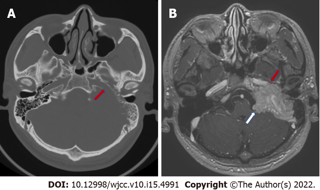
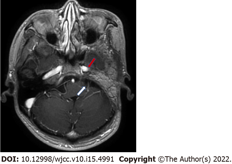
Figure 1 Preoperative computed tomography and magnetic resonance imaging images.
A: Computed tomography of the temporal bone showing the bone destruction of the horizontal segment of the internal carotid artery (red arrow); B: T1-weighted axial magnetic resonance imaging image showing the mass has partially wrapped around the horizontal internal carotid artery (red arrow) and has invaded the meninges and occupied the cerebral pontine area (white arrow).
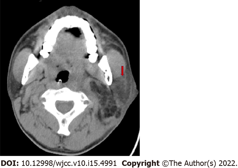
Figure 2 Postoperative neck computed tomography when swelling under the left ear and fever occurred.
A hypodensity shadow within the caudal lobe of the left parotid gland was present (red arrow).
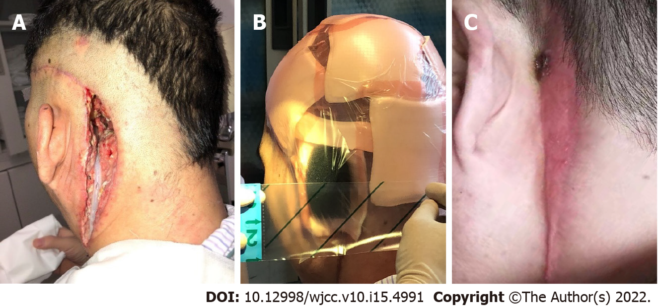
Figure 3 The operative wound.
A: The residual cavity of the operative wound after the debridement surgery and the placement of the drainage tube; B: VSD placement; C: The wound eventually healed after VSD had been performed.
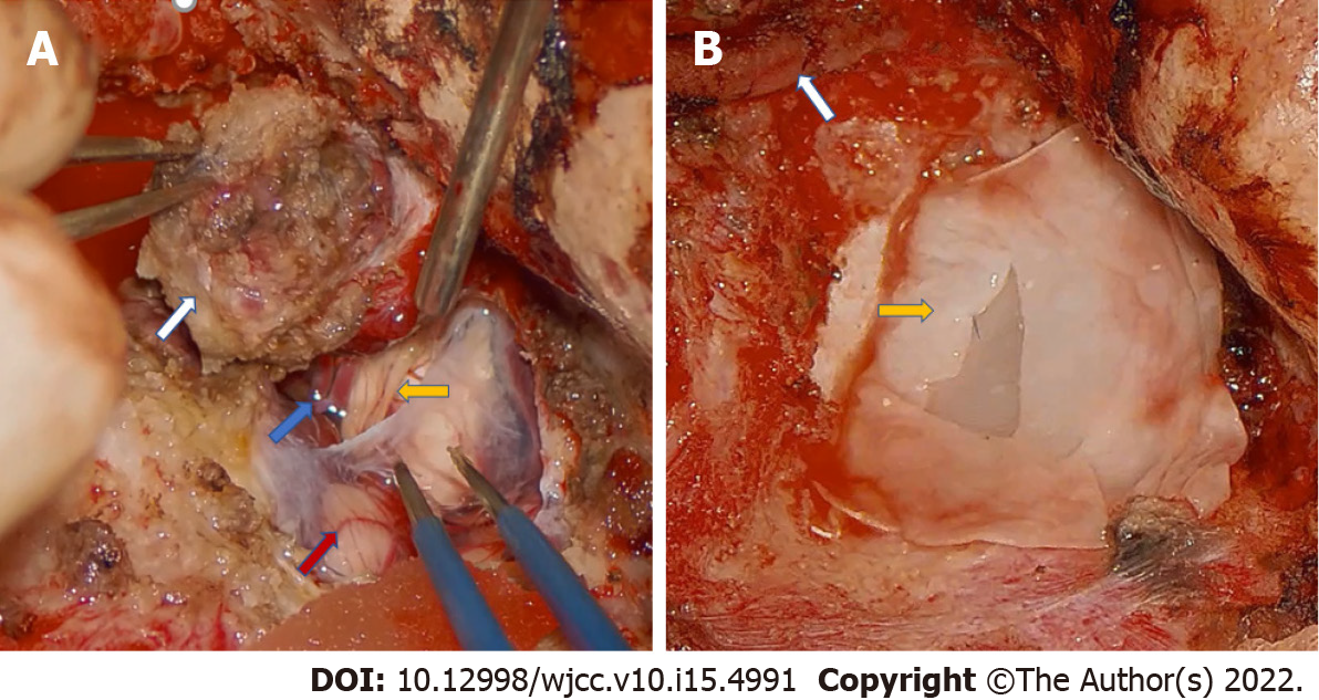
Figure 4 Intraoperative pictures of the second stage surgery.
A: The tumor (white arrow) that invaded the meninges; The anterior inferior cerebellar artery (blue arrow); The cochlear nerve (yellow arrow); The cerebellum (red arrow); B: The artificial dura mater (yellow arrow) used to repair the dura of the posterior fossa; The internal carotid artery (white arrow).
Figure 5 Enhanced temporal magnetic resonance imaging images at 6 mo after the second stage operation.
Infection or tumor recurrence was not found. The horizontal carotid artery (red arrow). The cerebral pontine area (white arrow).
- Citation: Zhao Y, Zhao Y, Zhang LQ, Feng GD. Postoperative infection of the skull base surgical site due to suppurative parotitis: A case report. World J Clin Cases 2022; 10(15): 4991-4997
- URL: https://www.wjgnet.com/2307-8960/full/v10/i15/4991.htm
- DOI: https://dx.doi.org/10.12998/wjcc.v10.i15.4991


