©The Author(s) 2022.
World J Clin Cases. May 26, 2022; 10(15): 4964-4970
Published online May 26, 2022. doi: 10.12998/wjcc.v10.i15.4964
Published online May 26, 2022. doi: 10.12998/wjcc.v10.i15.4964
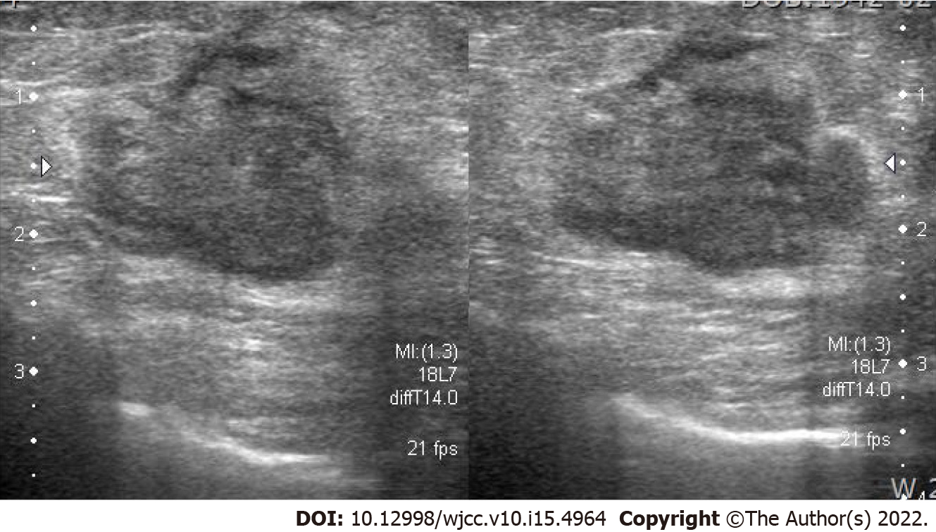
Figure 1 Ultrasonography revealed an ill-defined, lobulated, heterogeneous hypoechoic 2-cm mass on the left breast.
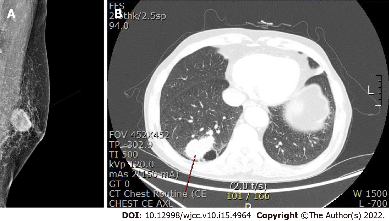
Figure 2 Computed tomography imaging.
A: Left-sided mammogram with a 3.2-cm oval, indistinct, hyperdense mass on the mid breast; B: A 5.1-cm lobulated mass in the posterior basal segment of the right lower lobe on chest computed tomography.
Figure 3 Metaplastic carcinoma of the breast.
A: The tumor is composed of pleomorphic and spindle cells with no ductal formation (hematoxylin and eosin, ×100); B: A necrotic focus is at the center of the tumor (arrow, hematoxylin and eosin, ×200); C: The spindle cells are positively stained for pancytokeratin (immunohistochemical staining, × 200).
Figure 4 Metastatic metaplastic carcinoma of the lung.
A: The metaplastic carcinoma (right side) is present in the lung parenchyma (left side) (hematoxylin and eosin, × 100); B and C: The pleomorphic tumor cells are epithelioid or spindle in shape with necrotic foci (hematoxylin and eosin, × 200).
- Citation: Kim HY, Lee S, Kim DI, Jung CS, Kim JY, Nam KJ, Choo KS, Jung YJ. Male metaplastic breast cancer with poor prognosis: A case report . World J Clin Cases 2022; 10(15): 4964-4970
- URL: https://www.wjgnet.com/2307-8960/full/v10/i15/4964.htm
- DOI: https://dx.doi.org/10.12998/wjcc.v10.i15.4964


