©The Author(s) 2022.
World J Clin Cases. May 16, 2022; 10(14): 4691-4697
Published online May 16, 2022. doi: 10.12998/wjcc.v10.i14.4691
Published online May 16, 2022. doi: 10.12998/wjcc.v10.i14.4691
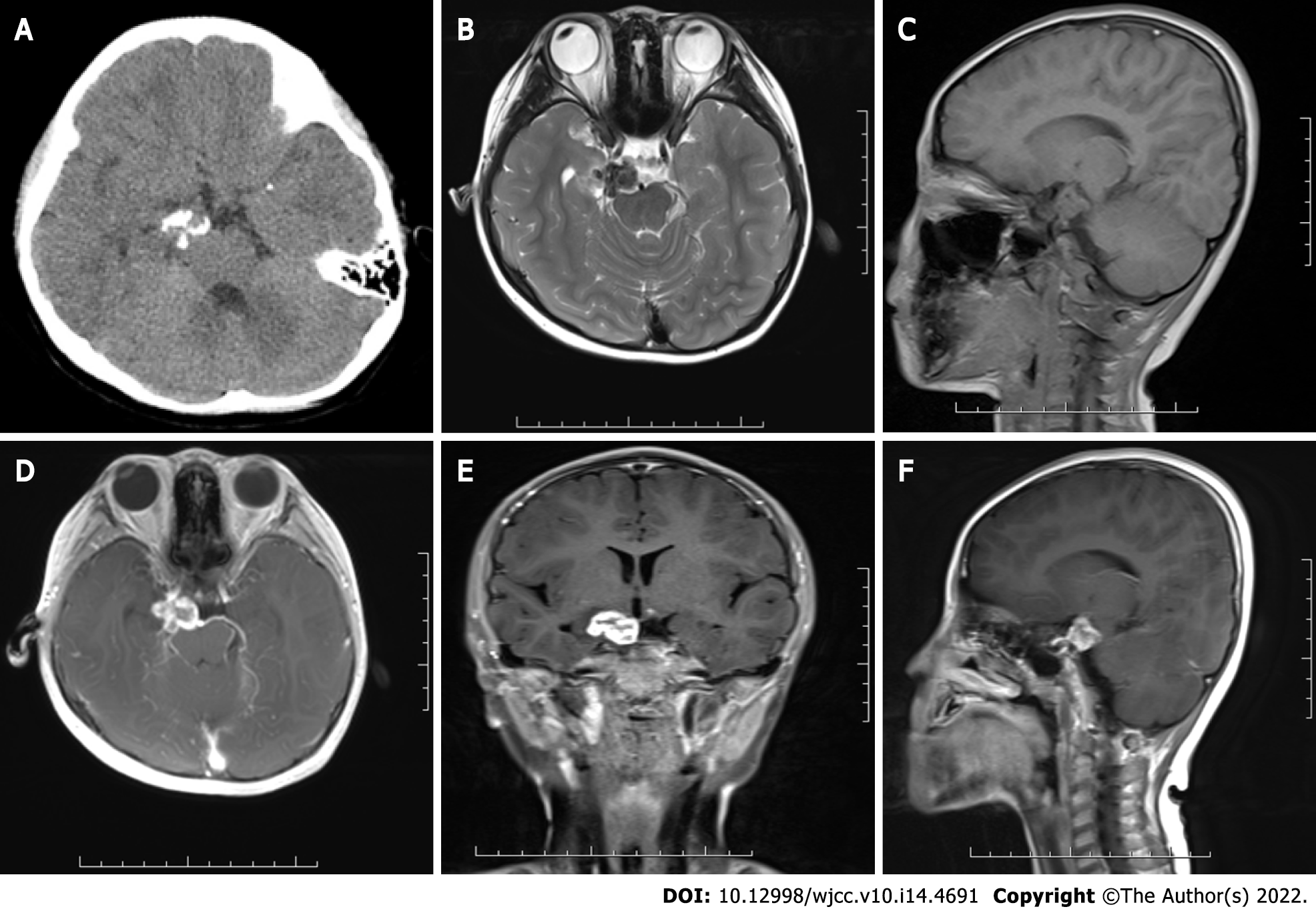
Figure 1 Preoperative cranial computed tomography image and magnetic resonance images.
A: Non-constrast computed tomography showed a mixed density mass in the suprasellar cistern with multiple patchy calcifications; B and C: Magnetic resonance images showed a lesion which appeared isointense on sagittal T1-weighted image and hypointense to isointense on axial T2-weighted image; D-F: Axial, coronal and sagittal contrast-enhanced T1-weighted images showed a ring-enhanced lesion.
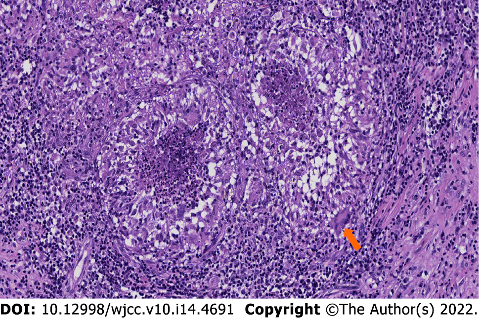
Figure 2 Histopathological findings.
The microscopic section showed epithelioid cell proliferation with caseous necrosis and infiltration of surrounding Langerhans giant cells (white arrow) and lymphocytes (× 100).
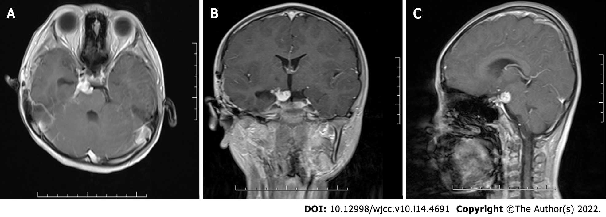
Figure 3 Postoperative cranial magnetic resonance images.
A and B: Axial and coronal contrast-enhanced T1-weighted images at 3 mo after surgery showed an enhanced residual lesion, which was significantly smaller than that before surgery; C: Sagittal image of the same examination showed that compression of the lesion on the pons and midbrain was reduced.
- Citation: Zhao BB, Tian C, Fu LJ, Zhang XB. Suprasellar cistern tuberculoma presenting as unilateral ocular motility disorder and ptosis: A case report. World J Clin Cases 2022; 10(14): 4691-4697
- URL: https://www.wjgnet.com/2307-8960/full/v10/i14/4691.htm
- DOI: https://dx.doi.org/10.12998/wjcc.v10.i14.4691


