©The Author(s) 2022.
World J Clin Cases. May 16, 2022; 10(14): 4676-4683
Published online May 16, 2022. doi: 10.12998/wjcc.v10.i14.4676
Published online May 16, 2022. doi: 10.12998/wjcc.v10.i14.4676
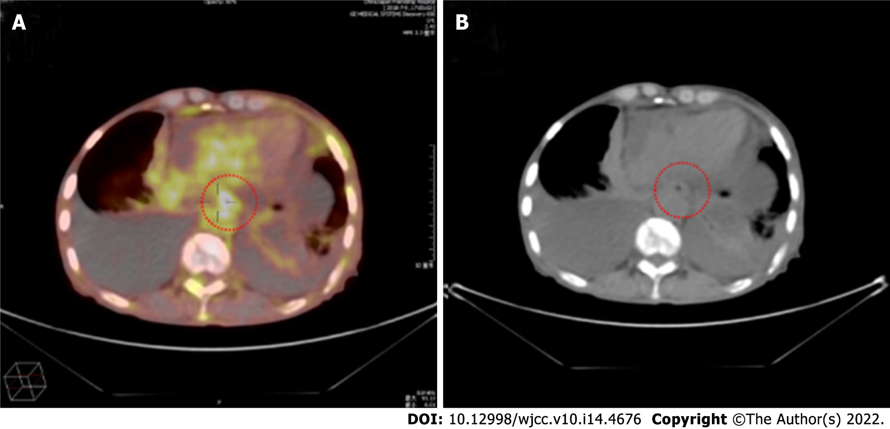
Figure 1 Positron emission tomography/computed tomography examination.
A, B: Diffuse thickening of the esophagus with an elevated standard uptake value and bilateral pleural effusion.
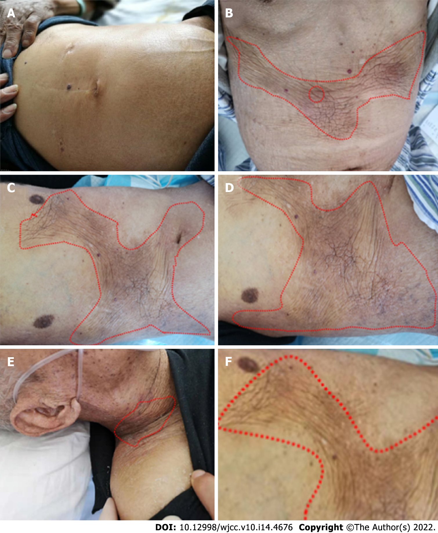
Figure 2 Progression of the cutaneous metastasis disease.
A: In July 2018 the normal abdominal wall skin at the beginning of diagnosed esophageal squamous cell carcinoma; B: In March 2019, the patient realized that the skin of the abdominal wall was hard and rough, and the color became darker than normal skin. At the same time, a fine needle biopsy of the skin of the changing part was performed; C: In early May 2019, the patient’s cutaneous metastasis significantly increased before the anterior chest wall; D: In early June 2019, the color of the skin lesions was lighter than before. The texture was soft, and the skin lesion did not expand; E, F: At the end of June, the patient’s cutaneous metastasis transferred to the anterior chest wall and abdominal wall as well as to the neck and lower limbs.
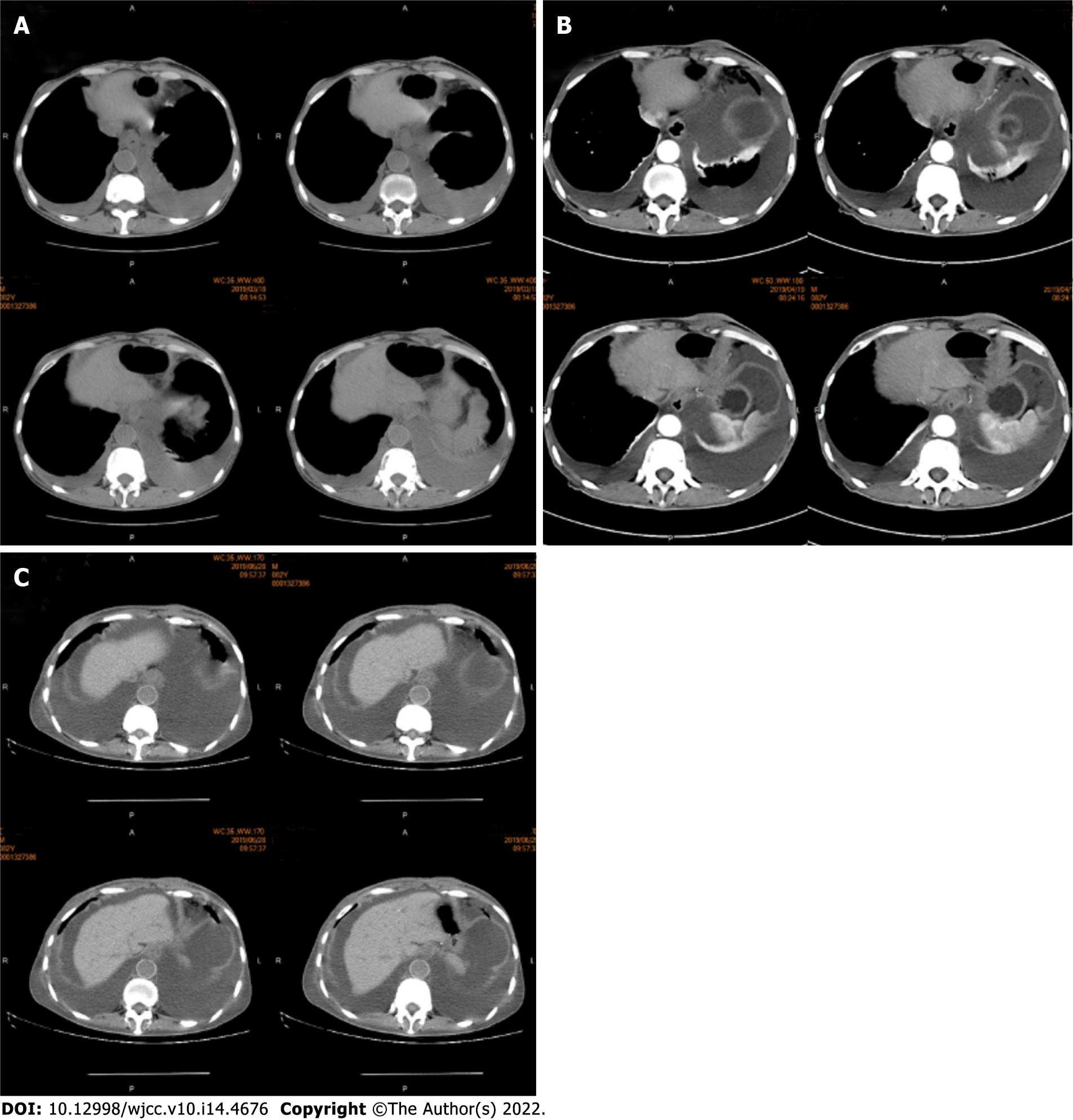
Figure 3 Abdominal computed tomography of the skin and subcutaneous soft tissue.
A: The chest computed tomography presented pleural effusion, suggesting that the disease recurred; B: This suggested ascites effusion as well as lower esophagus dilation and esophageal wall thickening; C: After two cycles of nivolumab treatment, the effusion increased, and the lower esophagus thickening became obvious. The abdominal computed tomography showed that the abdominal wall skin was thicker than the other parts, but no subcutaneous nodules could be observed. Imaging diagnosis can easily overlook skin thickening.
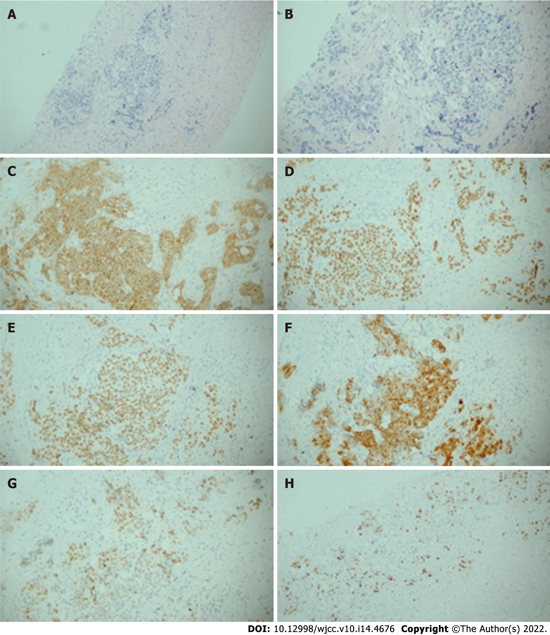
Figure 4 Histological patterns of skin lesions.
A, B: The hematoxylin and eosin staining of sites for the fine needle biopsy specimen revealed esophageal squamous cell carcinoma in A (magnification × 10), B (magnification × 20); C: Representative immunohistochemical staining for CK in the skin (magnification × 20); D:: Representative immunohistochemical staining for P40 in the skin (magnification × 20); E: Representative immunohistochemical staining for P63 in the skin (magnification × 20); F: Representative immunohistochemical staining for CK5/6 in the skin (magnification × 20); G: Representative immunohistochemical staining for P53 in the skin (magnification × 20); H: Representative immunohistochemical staining for Ki67 in the skin (magnification × 10).
- Citation: Zhang RY, Zhu SJ, Xue P, He SQ. Cutaneous metastasis from esophageal squamous cell carcinoma: A case report. World J Clin Cases 2022; 10(14): 4676-4683
- URL: https://www.wjgnet.com/2307-8960/full/v10/i14/4676.htm
- DOI: https://dx.doi.org/10.12998/wjcc.v10.i14.4676


