©The Author(s) 2022.
World J Clin Cases. May 16, 2022; 10(14): 4550-4562
Published online May 16, 2022. doi: 10.12998/wjcc.v10.i14.4550
Published online May 16, 2022. doi: 10.12998/wjcc.v10.i14.4550
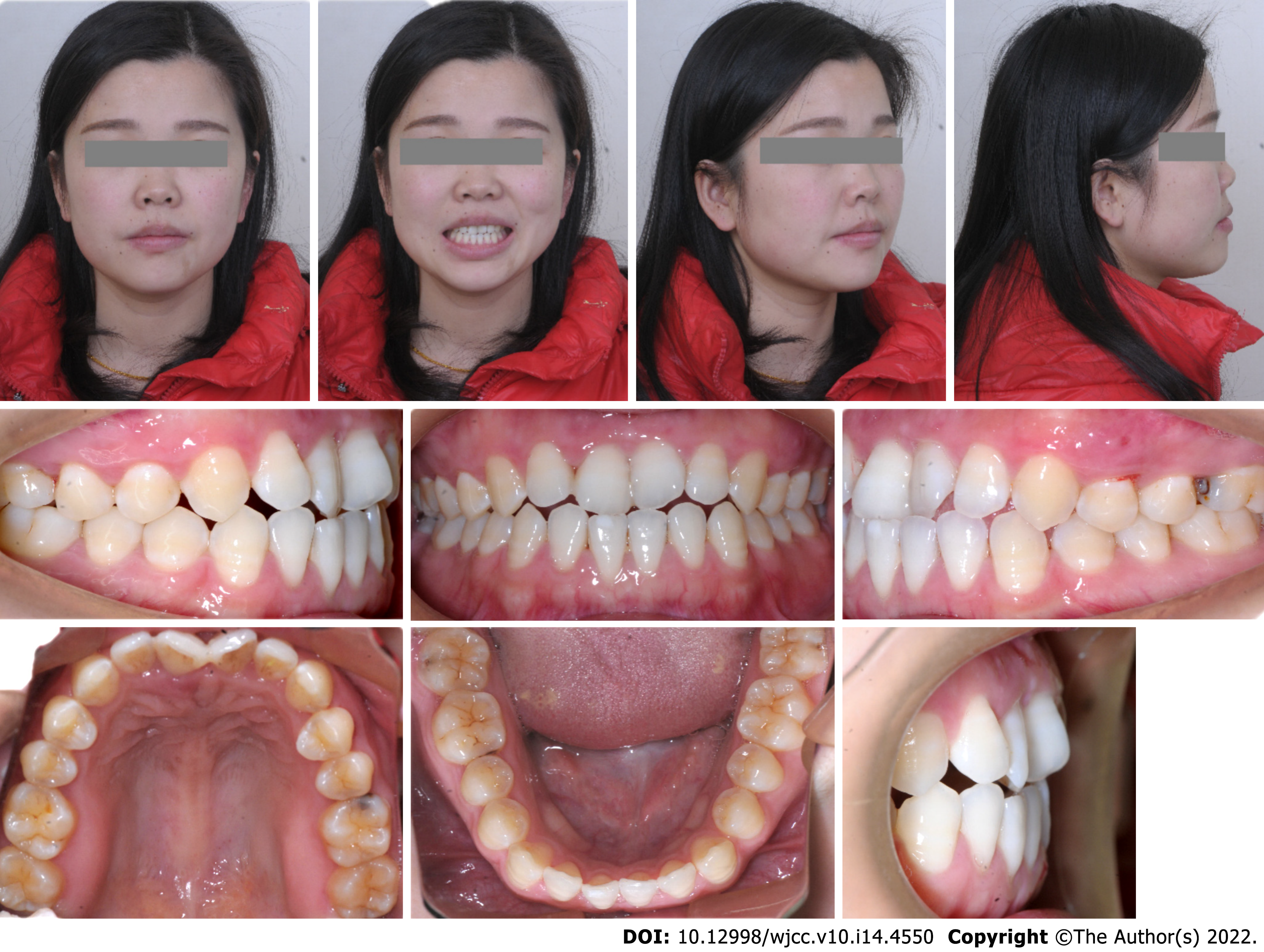
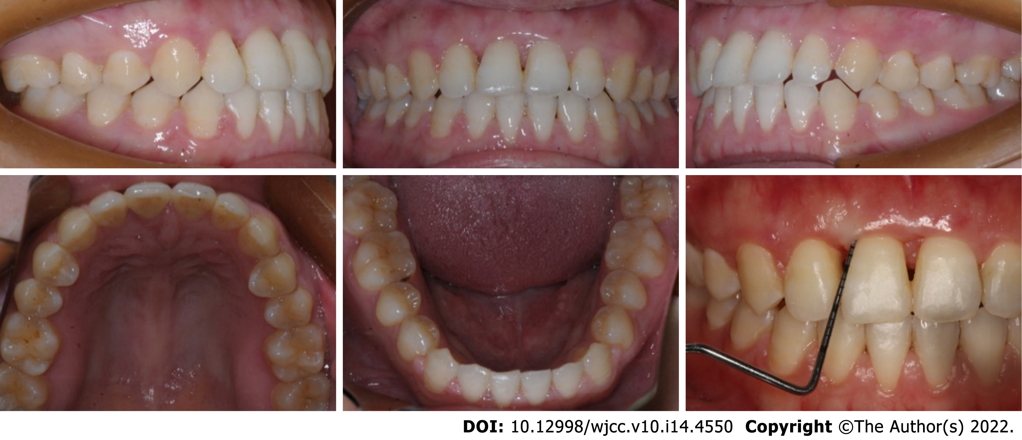
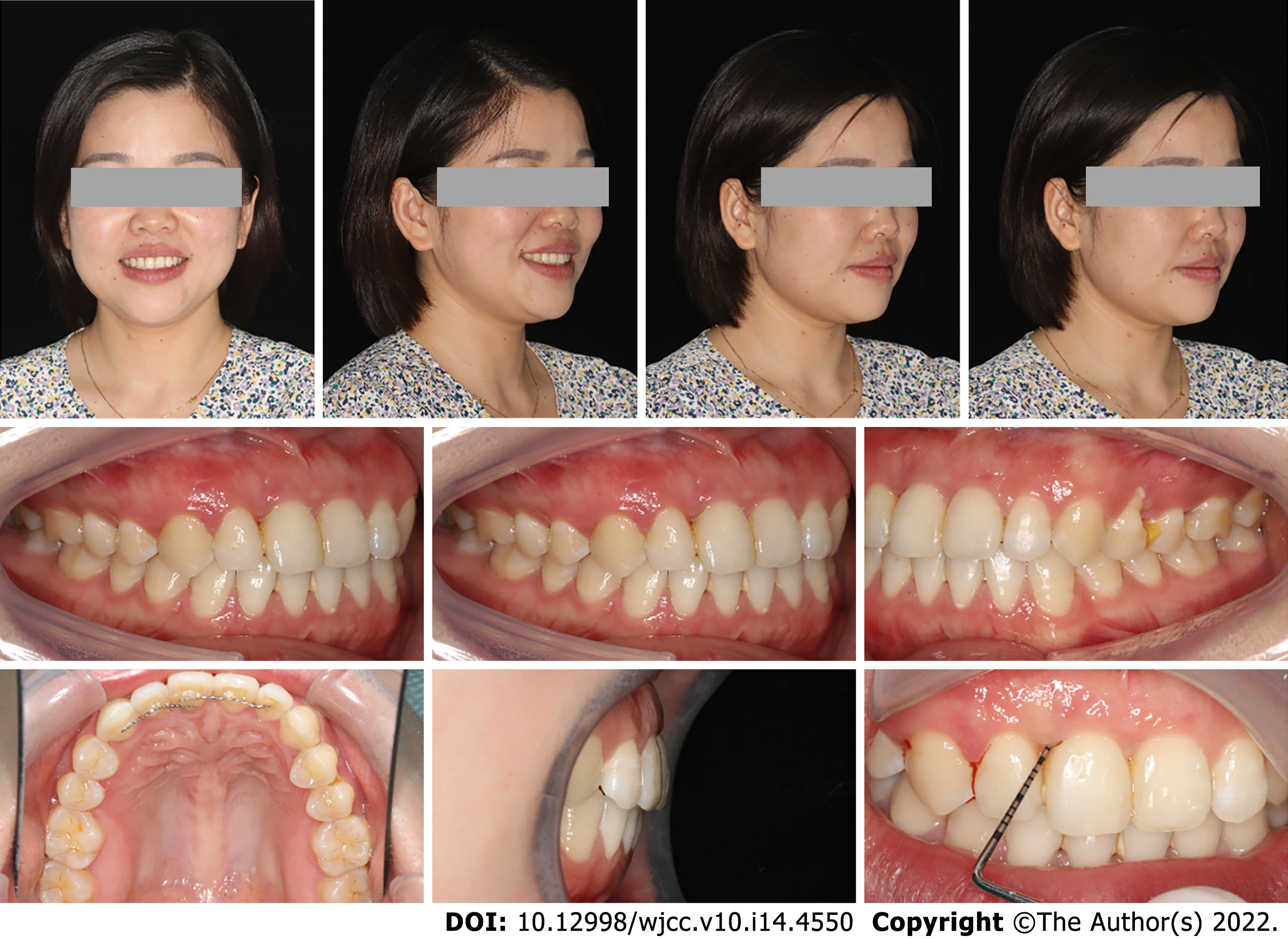
Figure 1 Extraoral and intraoral photographs before treatment.
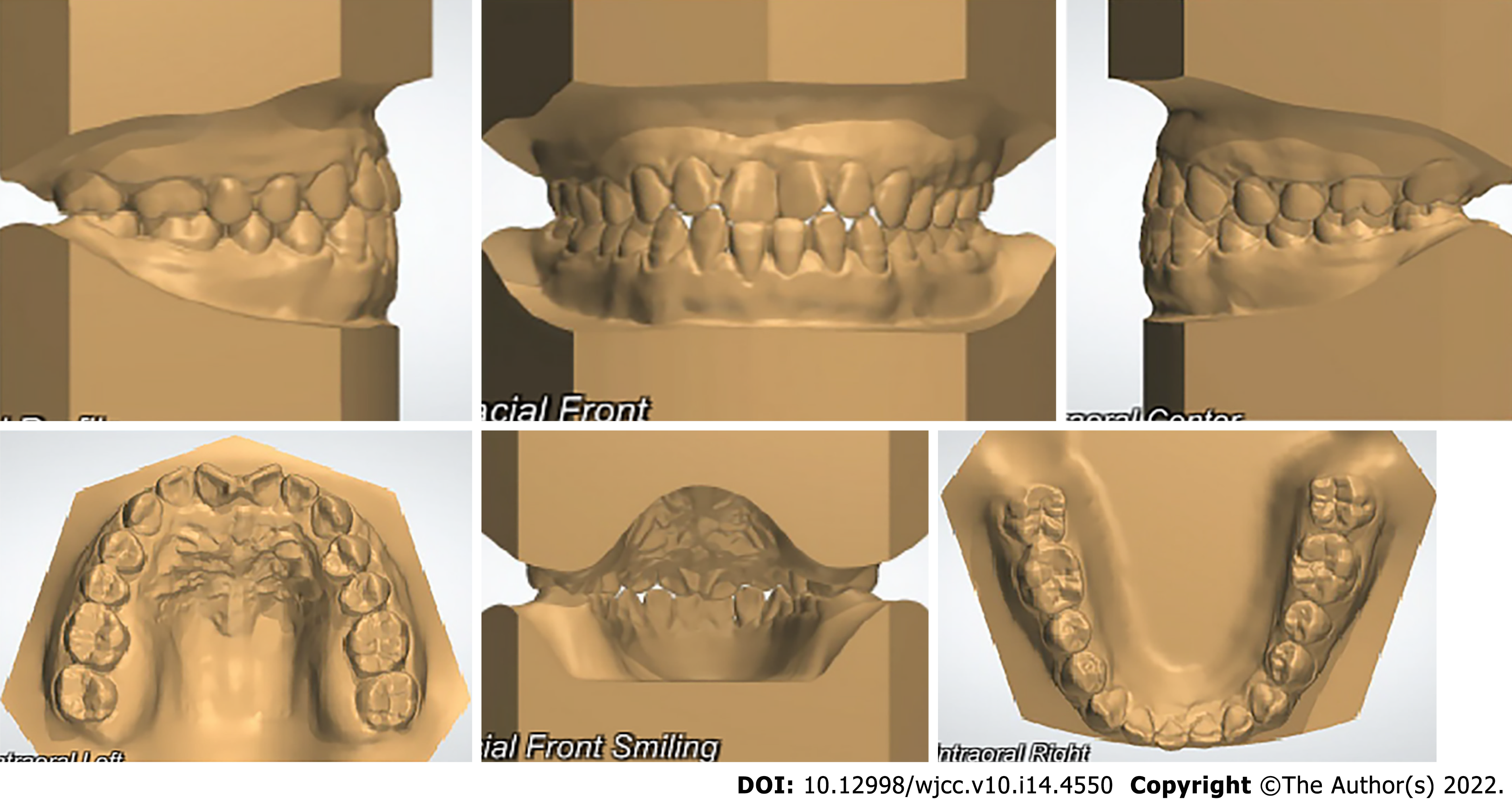
Figure 2 Pretreatment models.
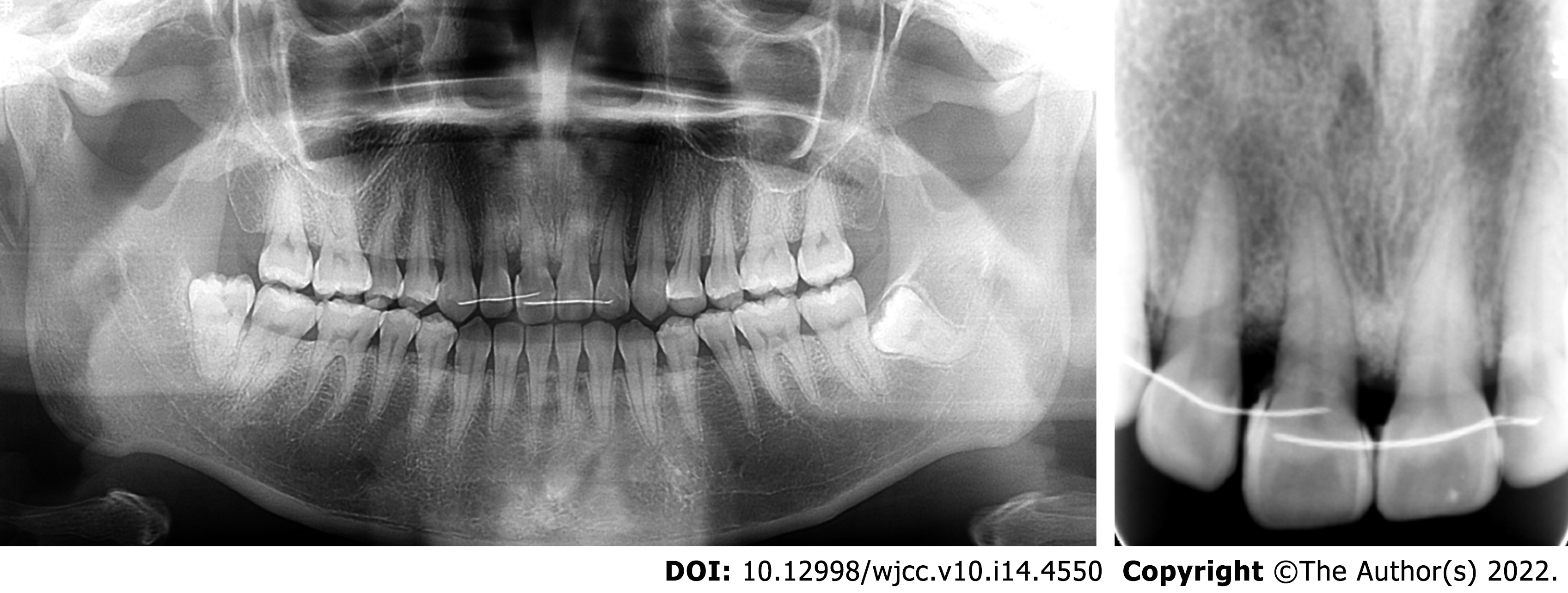
Figure 3 Pretreatment radiographs.
A: Panoramic radiograph; B: Apical radiograph; C: Cephalometric radiograph.
Figure 4 Diagram of the treatment process.
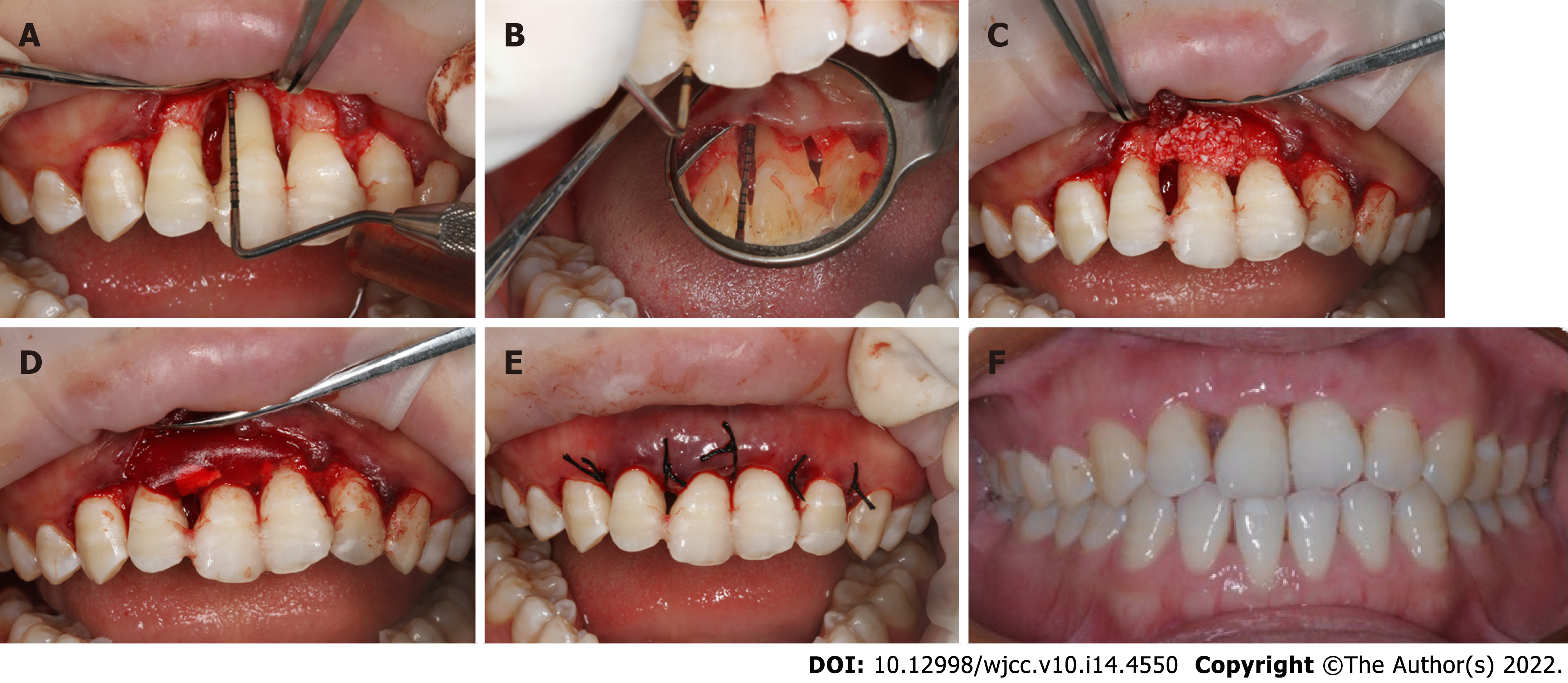
Figure 5 Periodontal surgery process and periodontal status 3 mo after surgery.
A and B: Facial and palatal bone defects after flap elevation; C and D: Bone particulate transplantation and collagen membrane insertion; E: Operation after suturing; F: Periodontal status 2 mo after surgery.
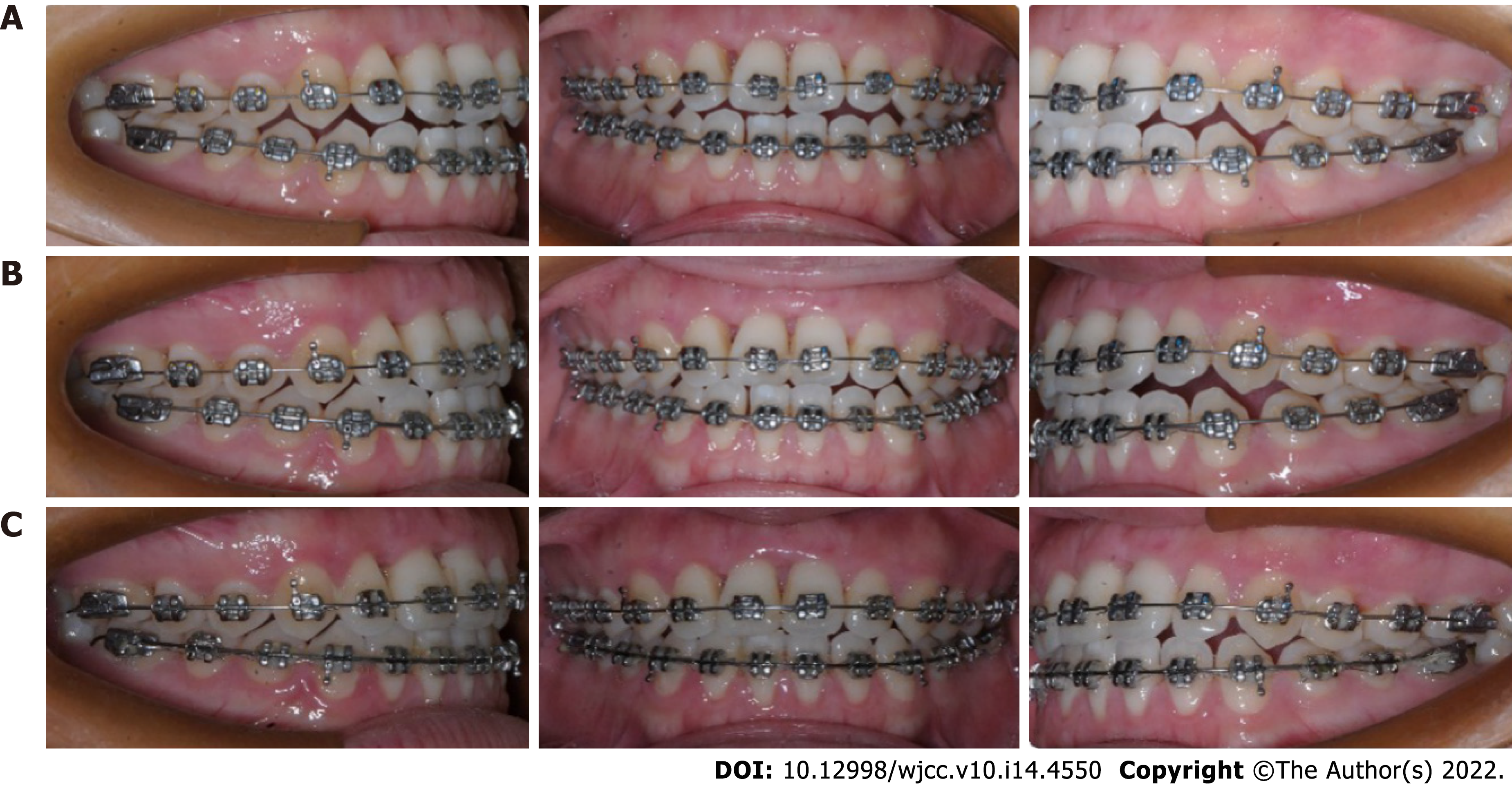
Figure 6 Treatment progress.
A-C: Photographs at 1, 2 and 3 mo after the start of orthodontic treatment.
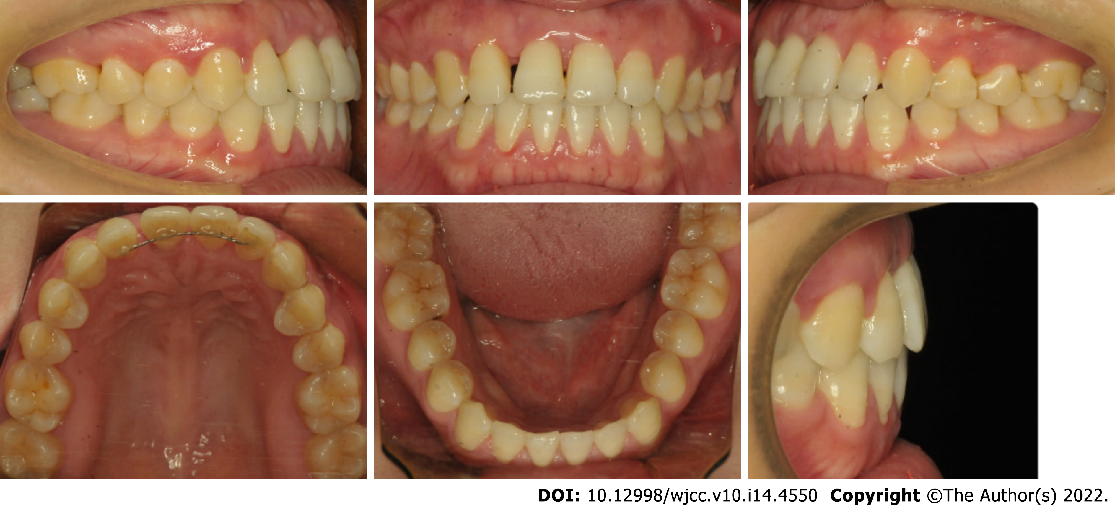
Figure 7 Posttreatment intraoral photographs and periodontal probing.
The periodontal tissue was in good condition, with no deep periodontal pockets.
Figure 8 Intraoral photographs after a follow-up period of 15 mo.
A space of 1 mm was observed between the upper right lateral incisor and the upper right central incisor.
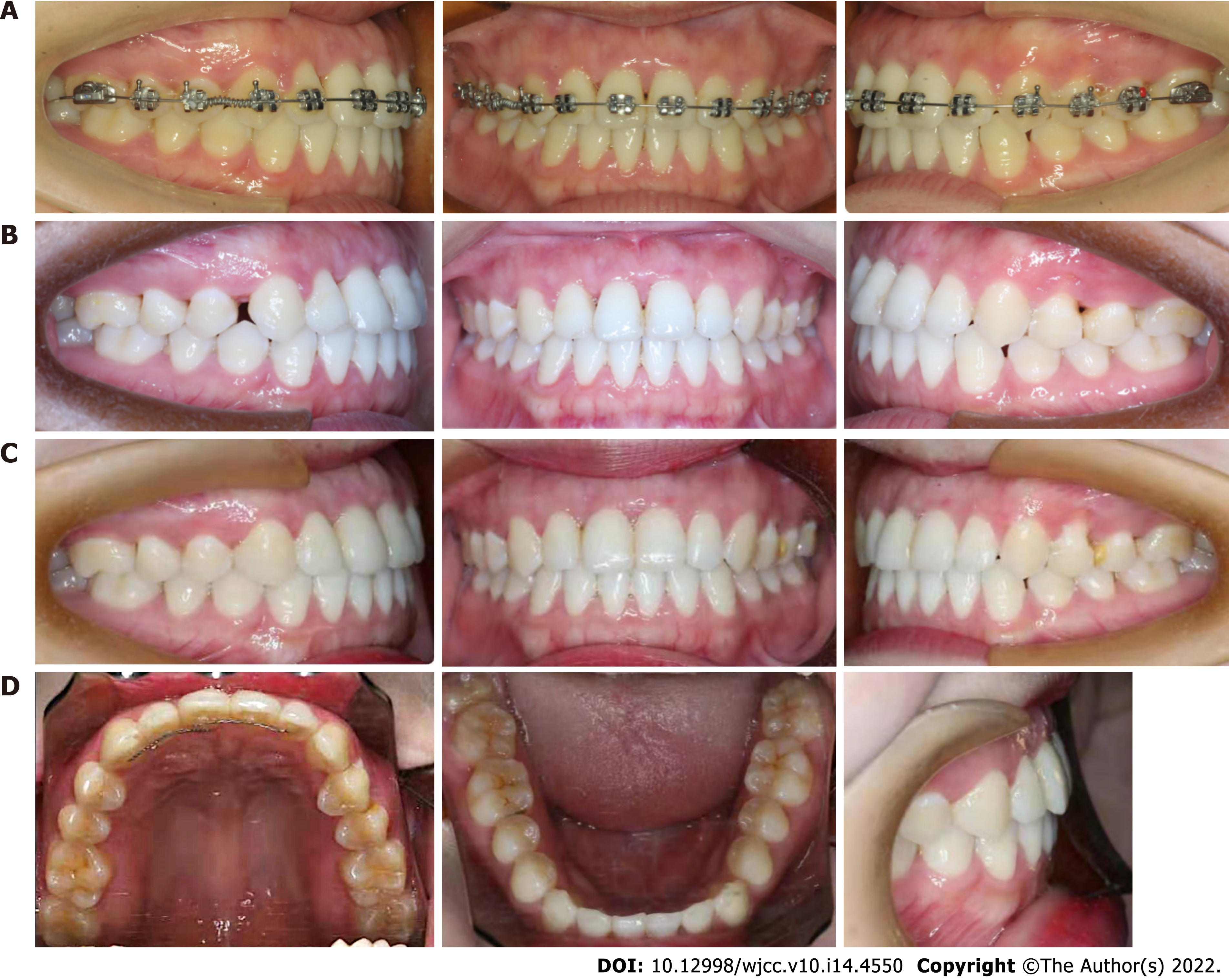
Figure 9 Second phase of orthodontic treatment and follow-up.
A and B: Orthodontic treatment to open space distal to the upper right canine; C: Treatment result.
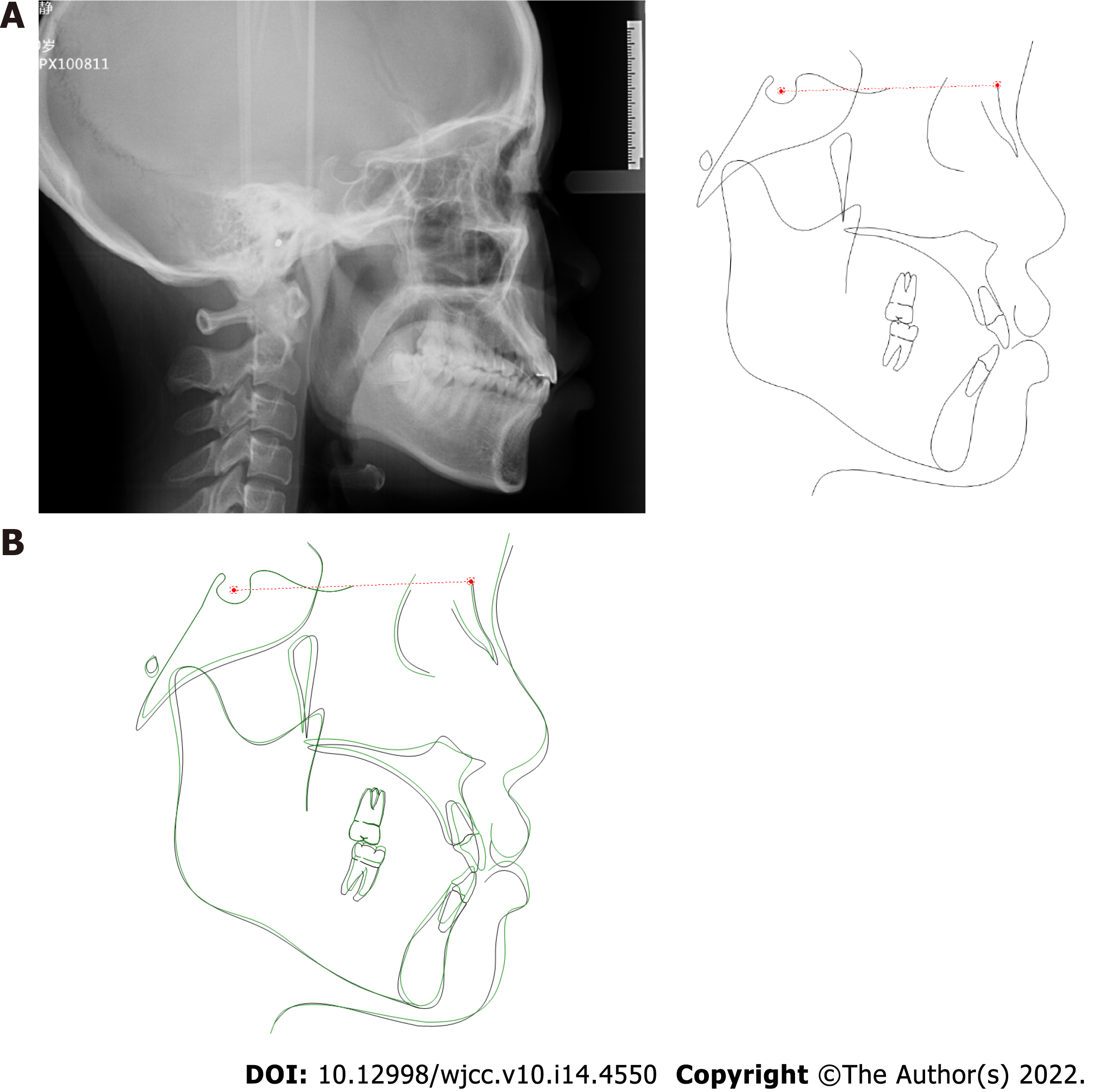
Figure 10 Posttreatment cephalometric radiograph.
A: Cephalometric radiograph after treatment; B: Conventional cephalometric superimposition of tracings before and after treatment.
Figure 11 Posttreatment radiographs (43 mo after periodontal surgery).
Figure 12 Posttreatment extraoral and intraoral photographs and periodontal probing on follow-up 1 year after orthodontic treatment.
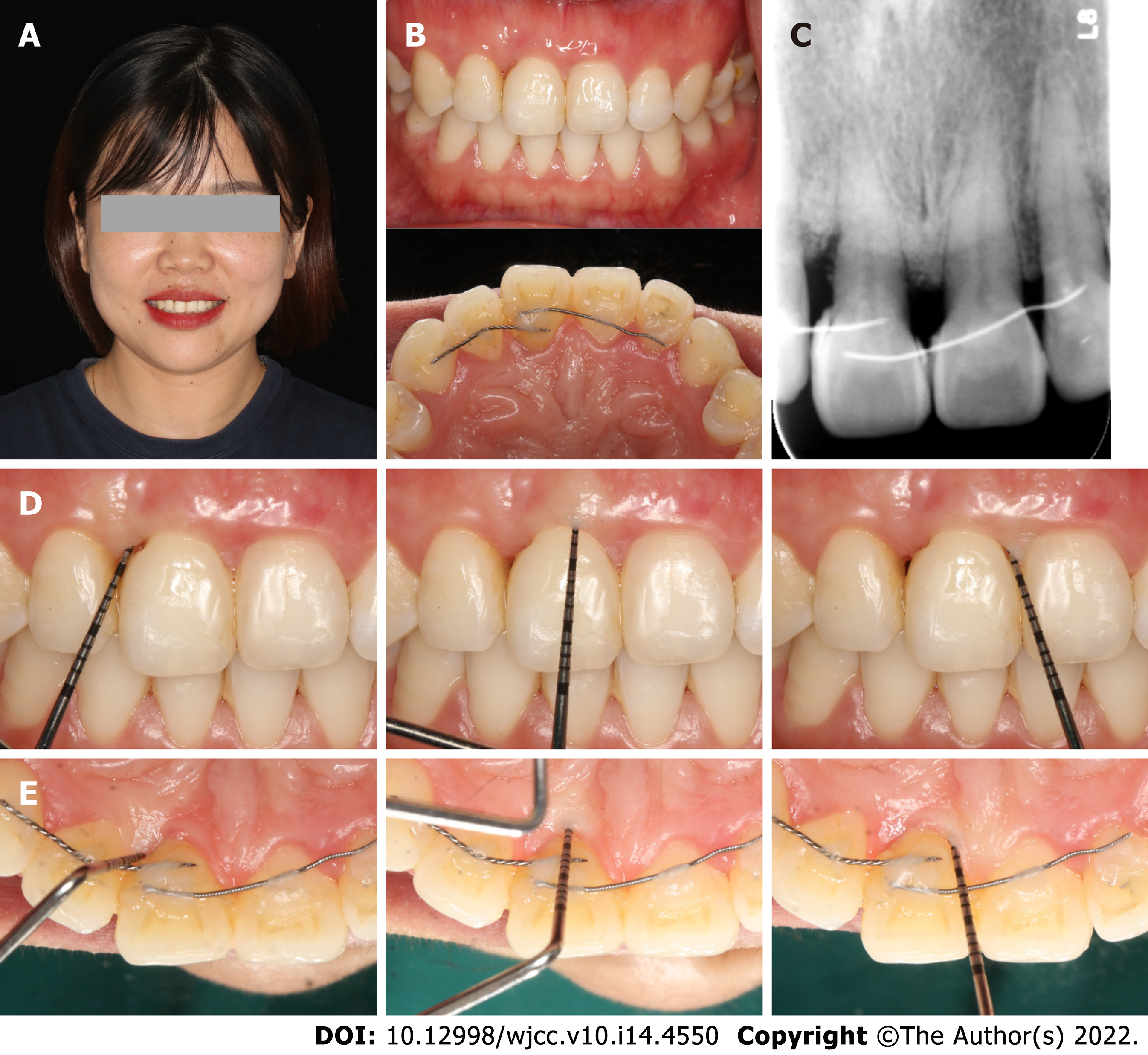
Figure 13 Follow-up 3 years after treatment.
A and B: Posttreatment extraoral and intraoral photographs; C: Apical radiograph; D and E: Periodontal probing.
- Citation: Jiang K, Jiang LS, Li HX, Lei L. Periodontal-orthodontic interdisciplinary management of a “periodontally hopeless” maxillary central incisor with severe mobility: A case report and review of literature. World J Clin Cases 2022; 10(14): 4550-4562
- URL: https://www.wjgnet.com/2307-8960/full/v10/i14/4550.htm
- DOI: https://dx.doi.org/10.12998/wjcc.v10.i14.4550


