©The Author(s) 2022.
World J Clin Cases. Apr 26, 2022; 10(12): 3849-3855
Published online Apr 26, 2022. doi: 10.12998/wjcc.v10.i12.3849
Published online Apr 26, 2022. doi: 10.12998/wjcc.v10.i12.3849
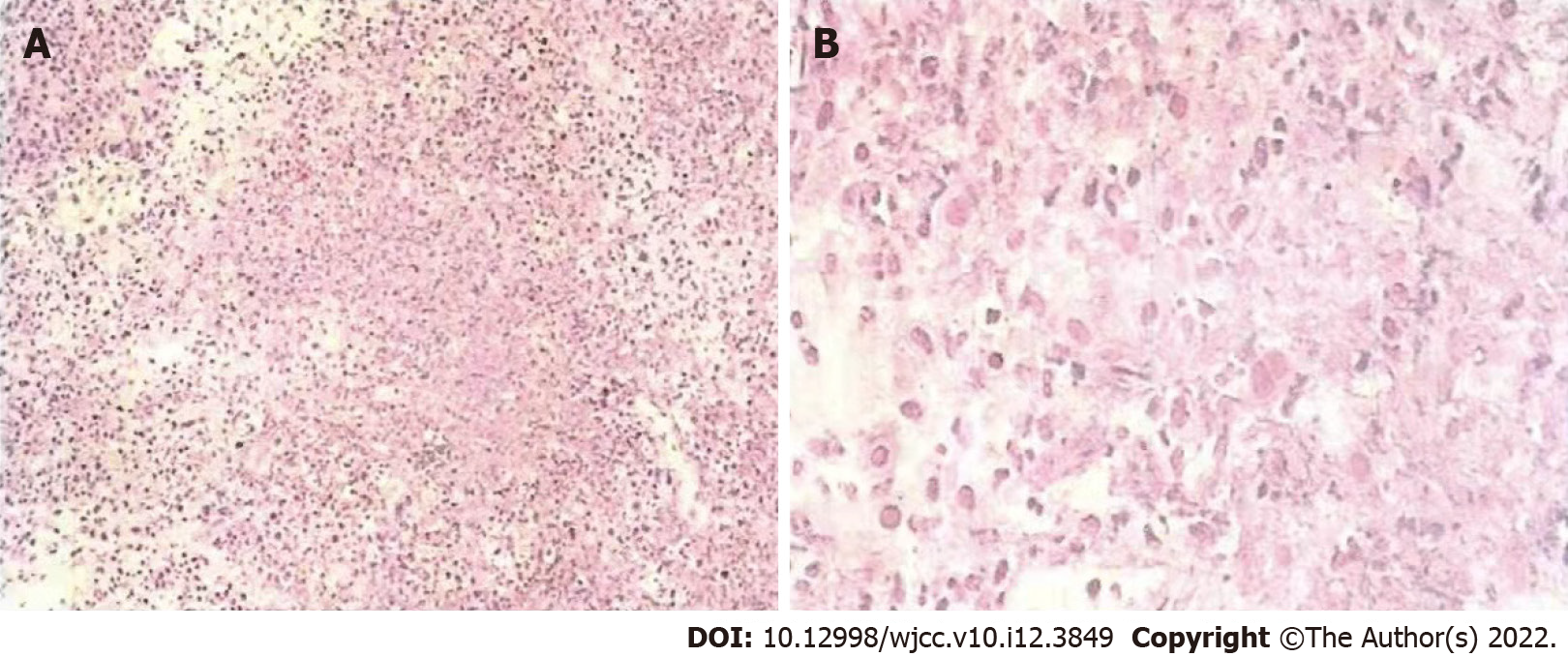
Figure 1 Pathological features.
A: First, the border was unclear, invasive growth was wide, and there was no capsule, and second, the tumor was extensively necrotic; B: Nuclear pleomorphism was evident, cells were small, and diffusely distributed, and finally, mitotic activity was high and invasion was strong.
Figure 2 Positron emission tomography-computed tomography showed left adrenal metastases (On September 14, 2017).
A: Computed tomography (CT) showed left adrenal metastasis; B: Cross-sectional positron emission tomography (PET) showed left adrenal metastasis; C: PET and CT fused layer showed left adrenal metastasis; D: Coronal PET-CT showed left adrenal metastasis.
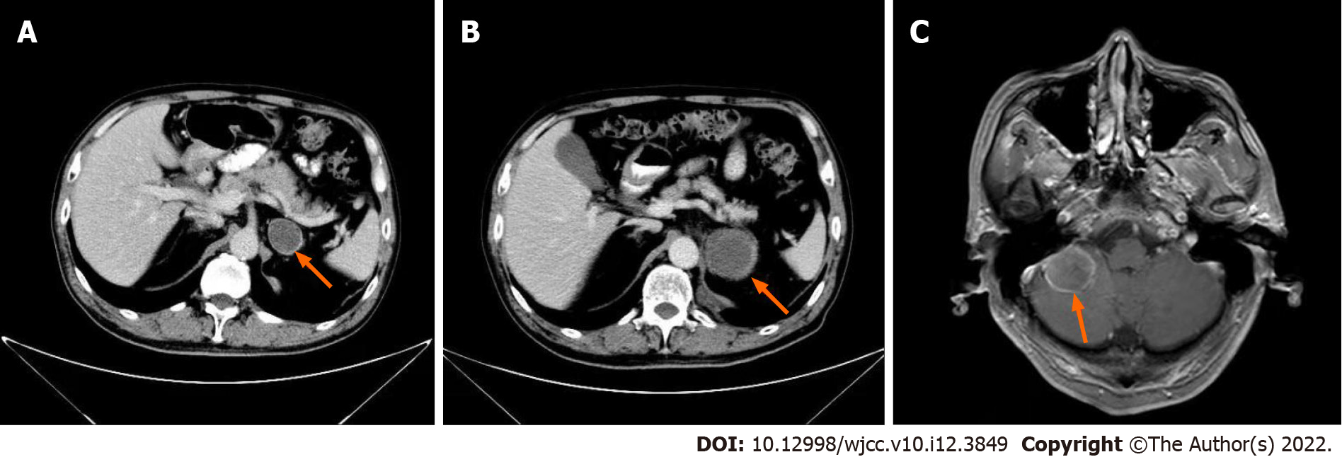
Figure 3 Metastasis during chemotherapy.
A: Left adrenal lesion after two cycles of TP chemotherapy; B: Significant enlargement of the left adrenal lesion after chemotherapy.C: Brain metastases.
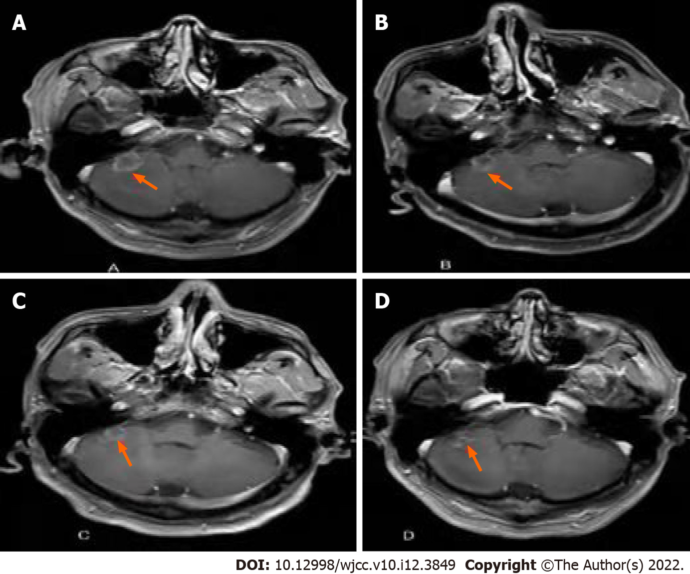
Figure 4 Changes in brain metastases during combined immune and targeted therapy.
A: Brain magnetic resonance imaging in October 2018; B: Brain magnetic resonance imaging in January 2019; C: Brain magnetic resonance imaging in June 2019; D: Brain magnetic resonance imaging in December 2019.
- Citation: Ma DX, Ding XP, Zhang C, Shi P. Combined targeted therapy and immunotherapy in anaplastic thyroid carcinoma with distant metastasis: A case report. World J Clin Cases 2022; 10(12): 3849-3855
- URL: https://www.wjgnet.com/2307-8960/full/v10/i12/3849.htm
- DOI: https://dx.doi.org/10.12998/wjcc.v10.i12.3849


