Copyright
©The Author(s) 2022.
World J Clin Cases. Apr 26, 2022; 10(12): 3754-3763
Published online Apr 26, 2022. doi: 10.12998/wjcc.v10.i12.3754
Published online Apr 26, 2022. doi: 10.12998/wjcc.v10.i12.3754
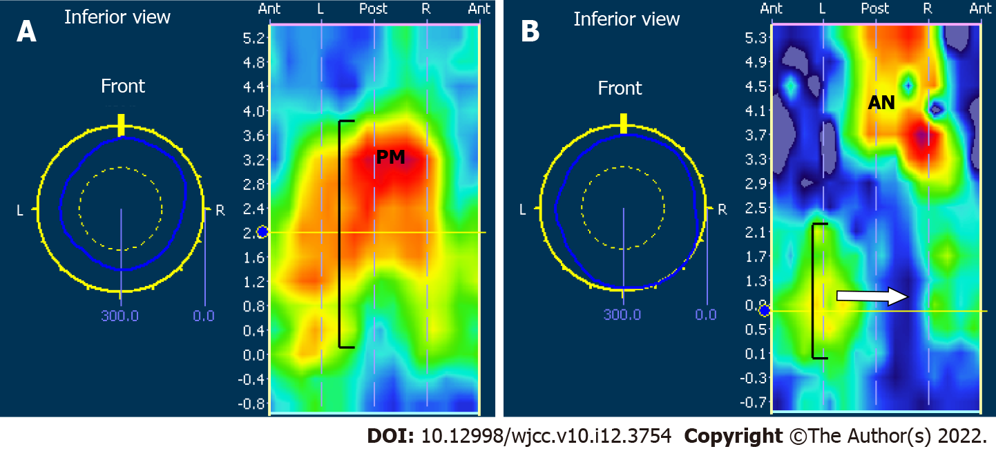
Figure 1 Three-dimensional profile map of three-dimensional high-resolution anorectal manometry in resting state before and after surgery (Dixon procedure) in a 55-year-old male patient with low rectal cancer who underwent neoadjuvant therapy.
A: Normal anal resting pressure was measured in this patient before surgery, and the tension of the puborectalis muscle is one important partial of the anal resting pressure; the length of the high pressure zone of the anal sphincter was normal in this case before surgery, which is marked with a brace ( [ ); B: After surgery, the patient’s anal resting pressure and length of the high pressure zone of the anal sphincter decreased significantly ( [ ). The blue area indicated with a white arrow is the defect area in the anal sphincter high-pressure zone; the high pressure zone (AN) was the manifestation of relative narrower anastomosis, which did not impact fecal passage.
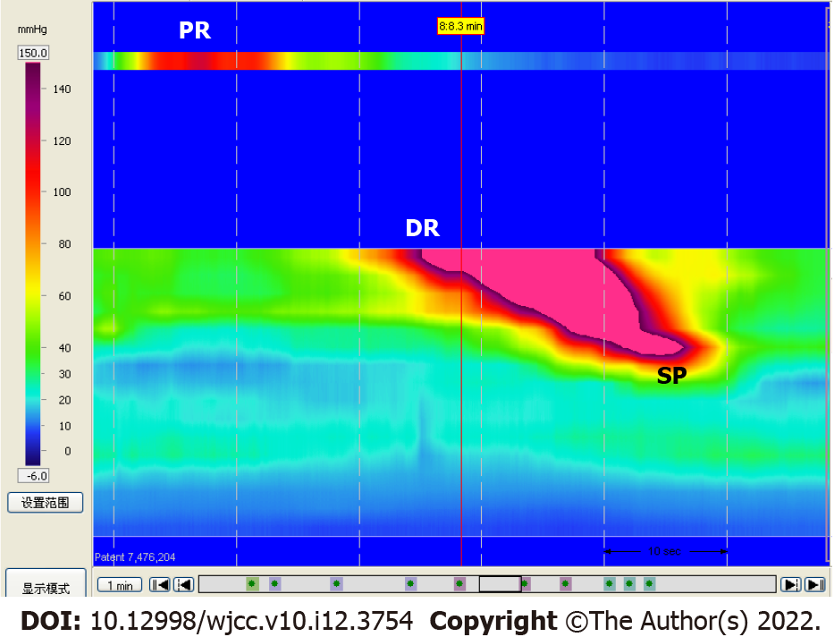
Figure 2 Two-dimensional map of three-dimensional high-resolution anorectal manometry in resting state in a patient with low rectal cancer after surgery (Dixon procedure).
The red area indicates involvement by spastic peristaltic neorectal contractions from the proximal to the distal end of the segment. Abscissa axis is the time intervals of 10 s between two white vertical dotted lines, and vertical axis indicates the position of electrode. The high pressure (a peristaltic contraction) initiated from the proximal segment of the new rectum at first 10 s time window (PR), which was detected by the electrode close to the balloon; about 10 s later, this spastic peristaltic contraction appeared in the distal rectum (DR) and then in the anal sphincter (SP), which were detected by the solid-state electrodes. DR: Distal rectum; SP: Sphincter; PR: Proximal rectum.
- Citation: Pi YN, Xiao Y, Wang ZF, Lin GL, Qiu HZ, Fang XC. Anorectal dysfunction in patients with mid-low rectal cancer after surgery: A pilot study with three-dimensional high-resolution manometry. World J Clin Cases 2022; 10(12): 3754-3763
- URL: https://www.wjgnet.com/2307-8960/full/v10/i12/3754.htm
- DOI: https://dx.doi.org/10.12998/wjcc.v10.i12.3754


