©The Author(s) 2022.
World J Clin Cases. Apr 16, 2022; 10(11): 3609-3614
Published online Apr 16, 2022. doi: 10.12998/wjcc.v10.i11.3609
Published online Apr 16, 2022. doi: 10.12998/wjcc.v10.i11.3609
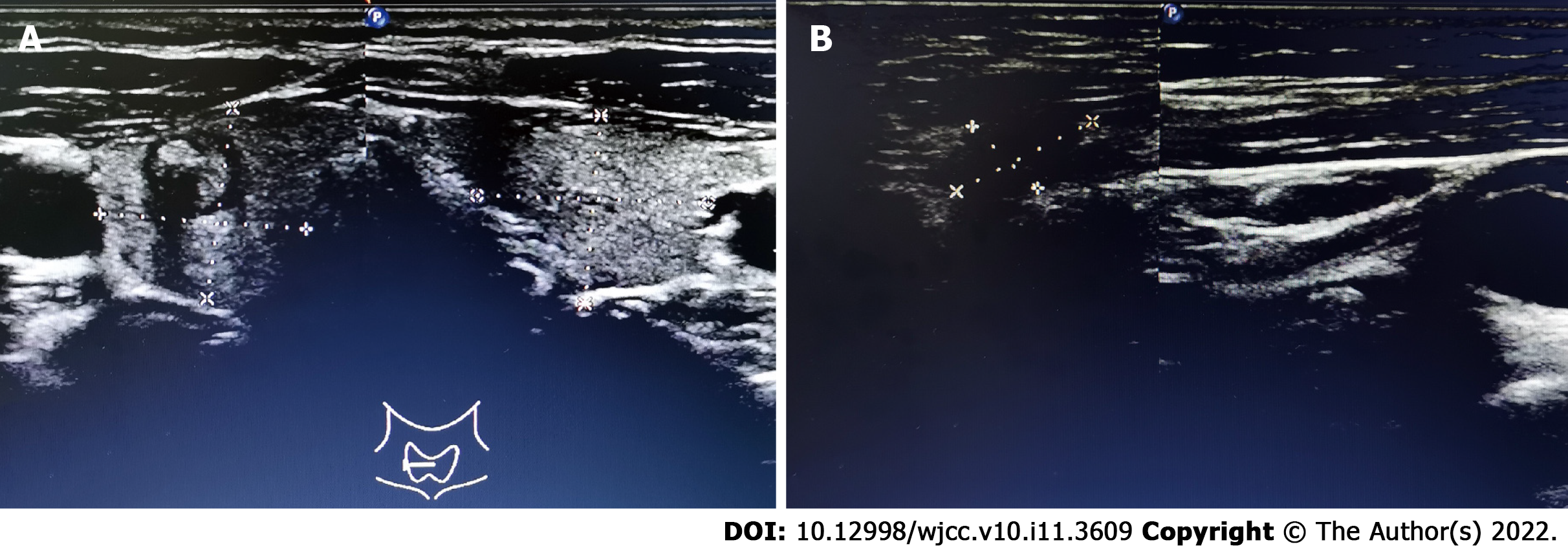
Figure 1 Ultrasound showed a nodule in the right thyroid lobe and an abnormal lymph node in left level V.
A: A 7.8 mm × 7.4 mm heterogeneous hypoechoic nodule with obscure boundary and hyperechoic punctuations was observed in the middle and upper part of the right lobe of the thyroid gland; B: A hypoechogenic structure could be detected in level V, which was approximately 14.0 mm × 7.0 mm in size, with irregular shape, obscure boundary, heterogeneous echo, and unclear lymphatic hilus.
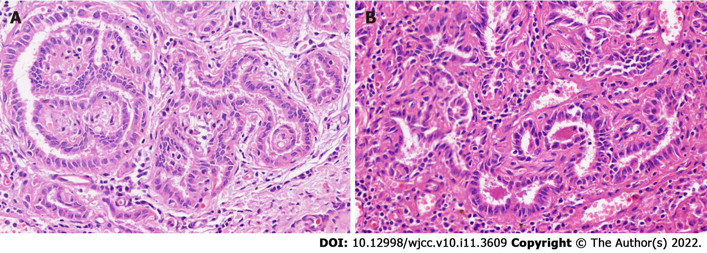
Figure 2 Hematoxylin and eosin staining of the right thyroid nodule and lymph node in left levels III and IV.
A: Papillary thyroid microcarcinoma in the right thyroid lobe; B: Lymph node metastasis in left levels III and IV. Magnification: 800 ×.
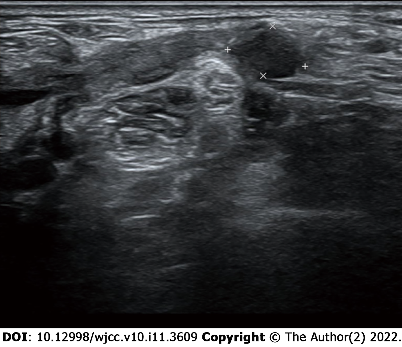
Figure 3 Ultrasound for the abnormal lymph nodes in the left supraclavicular and level V areas.
Several hypoechogenic structure were detected in the left supraclavicular and level V areas, one of which was approximately 10.1 mm × 6.5 mm in size with unclear lymphatic hilus.
- Citation: Ding M, Kong YH, Gu JH, Xie RL, Fei J. Papillary thyroid microcarcinoma with contralateral lymphatic skip metastasis and breast cancer: A case report. World J Clin Cases 2022; 10(11): 3609-3614
- URL: https://www.wjgnet.com/2307-8960/full/v10/i11/3609.htm
- DOI: https://dx.doi.org/10.12998/wjcc.v10.i11.3609


