©2013 Baishideng Publishing Group Co.
World J Clin Cases. Dec 16, 2013; 1(9): 295-297
Published online Dec 16, 2013. doi: 10.12998/wjcc.v1.i9.295
Published online Dec 16, 2013. doi: 10.12998/wjcc.v1.i9.295
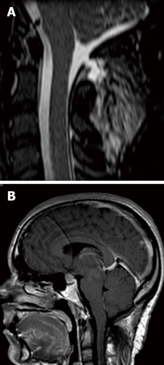
Figure 1 Magnetic resonance imaging signs of intracranial hypotension syndrome nine years after the craniectomy.
A: 7 mm × 2.5 mm dural defect with an extradural collection at the dorsal soft tissues of the cervical spine; B: Less prominent engorgement of the dural venous sinuses, further enlargement of the pituitary gland and download displacement or sagging of the brain with effacement of the perichiasmatic cisterns and the prepontine cistern.
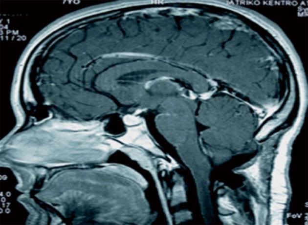
Figure 2 Magnetic resonance imaging signs of intracranial hypotension syndrome postoperatively.
There is significant engorgement of the dural venous sinuses and mild enlargement of the pituitary gland without signs of download displacement or sagging of the brain.
- Citation: Barkoula D, Bontozoglou N, Gatzonis S, Sakas D. Intracranial hypotension syndrome in a patient due to suboccipital craniectomy secondary to Chiari type malformation. World J Clin Cases 2013; 1(9): 295-297
- URL: https://www.wjgnet.com/2307-8960/full/v1/i9/295.htm
- DOI: https://dx.doi.org/10.12998/wjcc.v1.i9.295


