Copyright
©2013 Baishideng Publishing Group Co.
World J Clin Cases. Jul 16, 2013; 1(4): 152-154
Published online Jul 16, 2013. doi: 10.12998/wjcc.v1.i4.152
Published online Jul 16, 2013. doi: 10.12998/wjcc.v1.i4.152
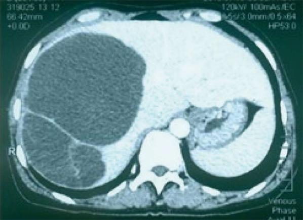
Figure 1 Computed tomography scan: A huge cystic mass with multiple septations is seen in the right lobe of the liver.
There was no enhancement in the cystic regions but the septa were enhanced and seemingly had calcification.
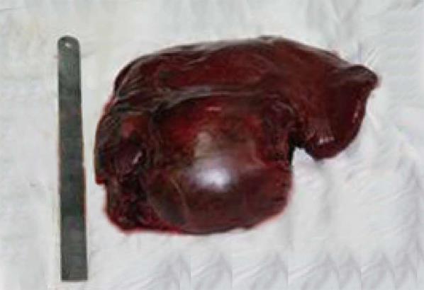
Figure 2 Gross sample: cystic mass in the right liver lobe, measuring 23 cm × 15 cm × 7 cm.
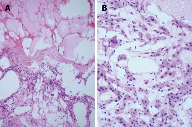
Figure 3 Histopathological findings.
A: Dilated lymphatic cavities accompanied with partial infarction. Lymph cells were seen lining the wall of the cyst, hematoxylin and eosin (HE), × 100; B: Simple squamous epithelia and eosinophilic hepatic cells (HE, × 200).
- Citation: Zhang YZ, Ye YS, Tian L, Li W. Rare case of a solitary huge hepatic cystic lymphangioma. World J Clin Cases 2013; 1(4): 152-154
- URL: https://www.wjgnet.com/2307-8960/full/v1/i4/152.htm
- DOI: https://dx.doi.org/10.12998/wjcc.v1.i4.152


