Copyright
©2013 Baishideng Publishing Group Co.
World J Clin Cases. Jun 16, 2013; 1(3): 96-105
Published online Jun 16, 2013. doi: 10.12998/wjcc.v1.i3.96
Published online Jun 16, 2013. doi: 10.12998/wjcc.v1.i3.96
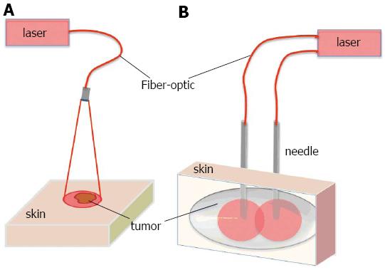
Figure 1 Representation for light delivery during surface and interstitial photodynamic therapy.
A: Surface illumination photodynamic therapy (PDT) for treating superficial malignancies. Laser light is directed to tissue surface via micro-lens fiber. Tumor is located superficially; B: Interstitial PDT treatment for deeper and thicker malignancies. Individual fibers are placed inside 19-gauge needles and inserted into tissue. Number of fibers is selected according to treated volume.
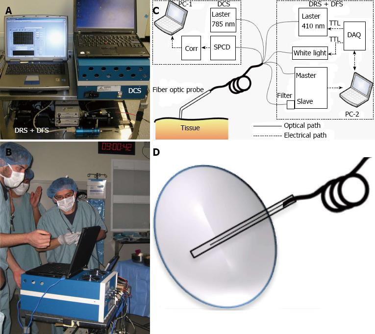
Figure 2 Clinical multi-modal optical instrument for photodynamic therapy dosimetry and response monitoring.
A: Picture of multi-modal clinical optical instrument; B: During the measurements at the operating room; C: Diagram of multi-modal clinical optical instrument; D: Interstitial optical probe for measurements in deep and thick tumors. Adapted from the reference[45] with the permission.
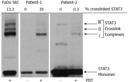
Figure 3 Signal transducer and activator of transcription 3 crosslinking as a molecular marker for local photodynamic therapy dose.
Signal transducer and activator of transcription 3 (STAT3) crosslinking for Patient-1 and Patient-2 with a human hypopharyngeal carcinoma cell line (FaDu) shown as a control. Adapted from the reference[74] with the permission. PDT: Photodynamic therapy.
- Citation: Sunar U. Monitoring photodynamic therapy of head and neck malignancies with optical spectroscopies. World J Clin Cases 2013; 1(3): 96-105
- URL: https://www.wjgnet.com/2307-8960/full/v1/i3/96.htm
- DOI: https://dx.doi.org/10.12998/wjcc.v1.i3.96


