Copyright
©2013 Baishideng.
World J Clin Cases. May 16, 2013; 1(2): 87-91
Published online May 16, 2013. doi: 10.12998/wjcc.v1.i2.87
Published online May 16, 2013. doi: 10.12998/wjcc.v1.i2.87
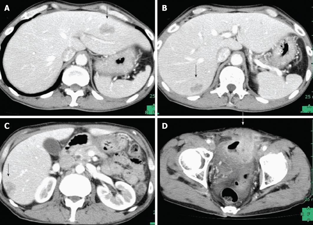
Figure 1 Computed tomography before treatment showed multiple liver metastases (arrows) and a primary upper rectal tumor that had invaded the urinary bladder.
A, B: Multiple liver metastases; C, D: A primary upper rectal tumor.
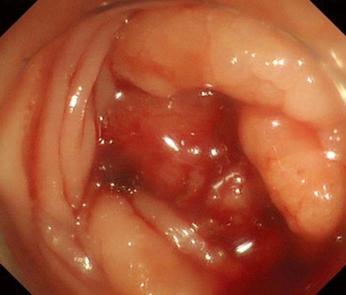
Figure 2 Colonoscopy revealed a circumferential lesion in the upper rectum and the lumen was almost completely obstructed.
Figure 3 On esophagogastroscopy, a depressed lesion with a little surrounding elevation was observed in the mid-thoracic esophagus, and biopsy resulted in a diagnosis of squamous cell carcinoma.
A: Esophagogastroscopy before treatment revealed a shallow depressed area in the middle third of the thoracic esophagus; B: After 6 cycles of chemotherapy, the esophageal lesion had flattened slightly, and there was good disease control.
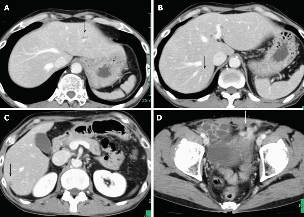
Figure 4 The computed tomography findings after 6 cycles of mFOLFOX6 plus cetuximab chemotherapy showed a good response in both the liver metastases (arrows) and primary lesion.
A, B: Liver metastases; C, D: Primary lesion.
- Citation: Utsunomiya S, Uehara K, Kurimoto T, Hirose K, Fukaya M, Takahashi Y, Taguchi Y, Itatsu K, Nagino M. Synchronous rectal and esophageal cancer treated with chemotherapy followed by two-stage resection. World J Clin Cases 2013; 1(2): 87-91
- URL: https://www.wjgnet.com/2307-8960/full/v1/i2/87.htm
- DOI: https://dx.doi.org/10.12998/wjcc.v1.i2.87


