©The Author(s) 2021.
World J Crit Care Med. Nov 9, 2021; 10(6): 323-333
Published online Nov 9, 2021. doi: 10.5492/wjccm.v10.i6.323
Published online Nov 9, 2021. doi: 10.5492/wjccm.v10.i6.323
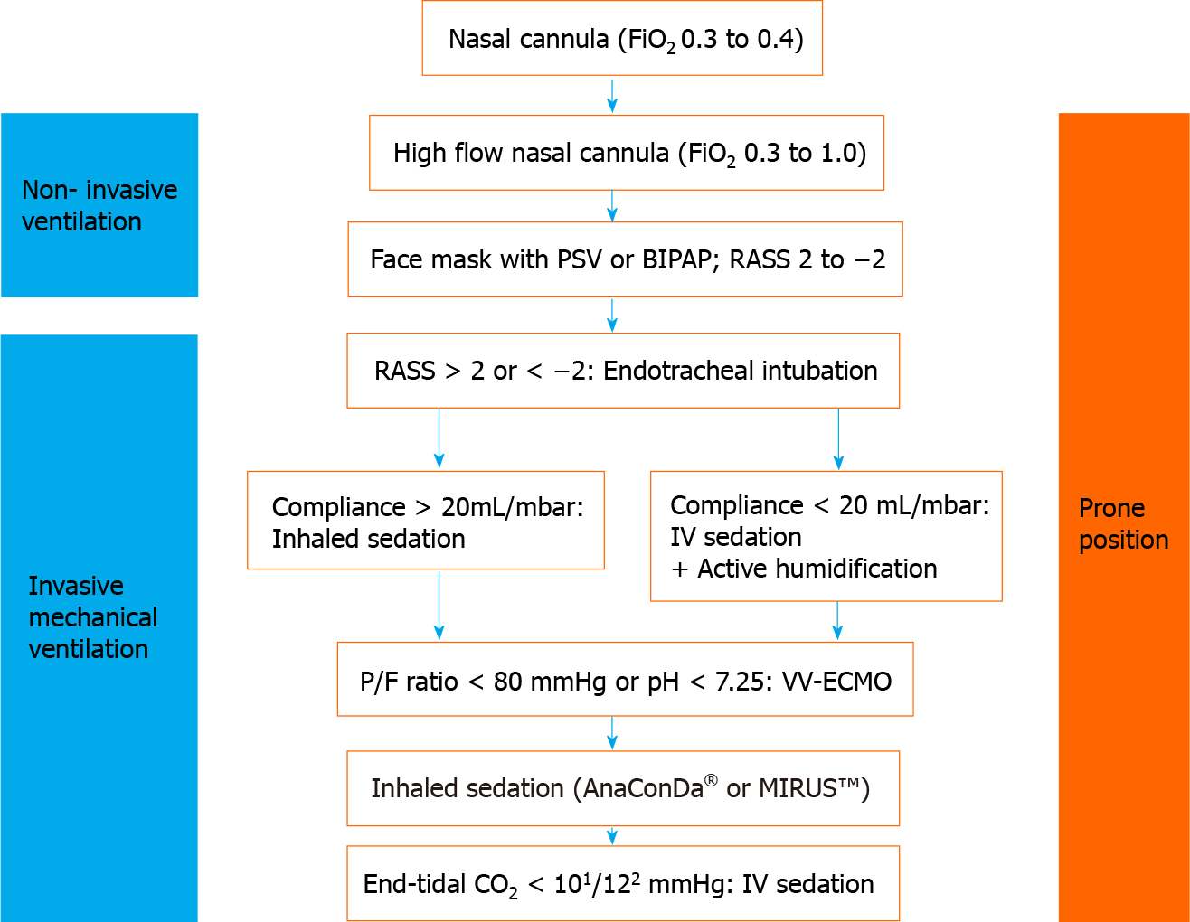
Figure 1 Overview of respiratory management of coronavirus disease 2019 related acute respiratory distress syndrome and inhaled sedation.
1AnaConDa®; 2MIRUSTM. RASS: Richmond Agitation-Sedation Scale; PSV: Pressure support ventilation; BIPAP: Bilevel positive airway pressure; FiO2: Fraction of inspired oxygen; PaO2: Partial pressure of oxygen; P/F-ratio: PaO2/FiO2; VV-ECMO: Veno-venous ECMO.
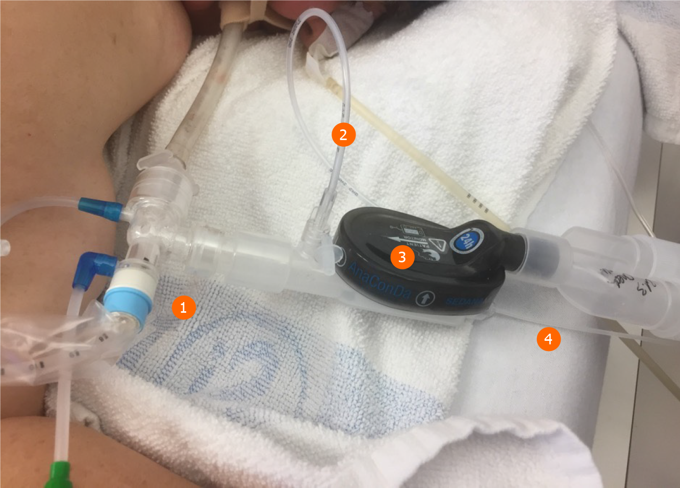
Figure 2 AnaConDa-S® system set up in prone position.
1Closed loop suction system; 2Port to monitor the volatile anesthetic and CO2; 3AnaConDa®-S with anesthesia gas reflector, bacterial and viral filter, and heat and moisture exchanger; 4Evaporator with liquid line from syringe pump and liquid isoflurane or sevoflurane.
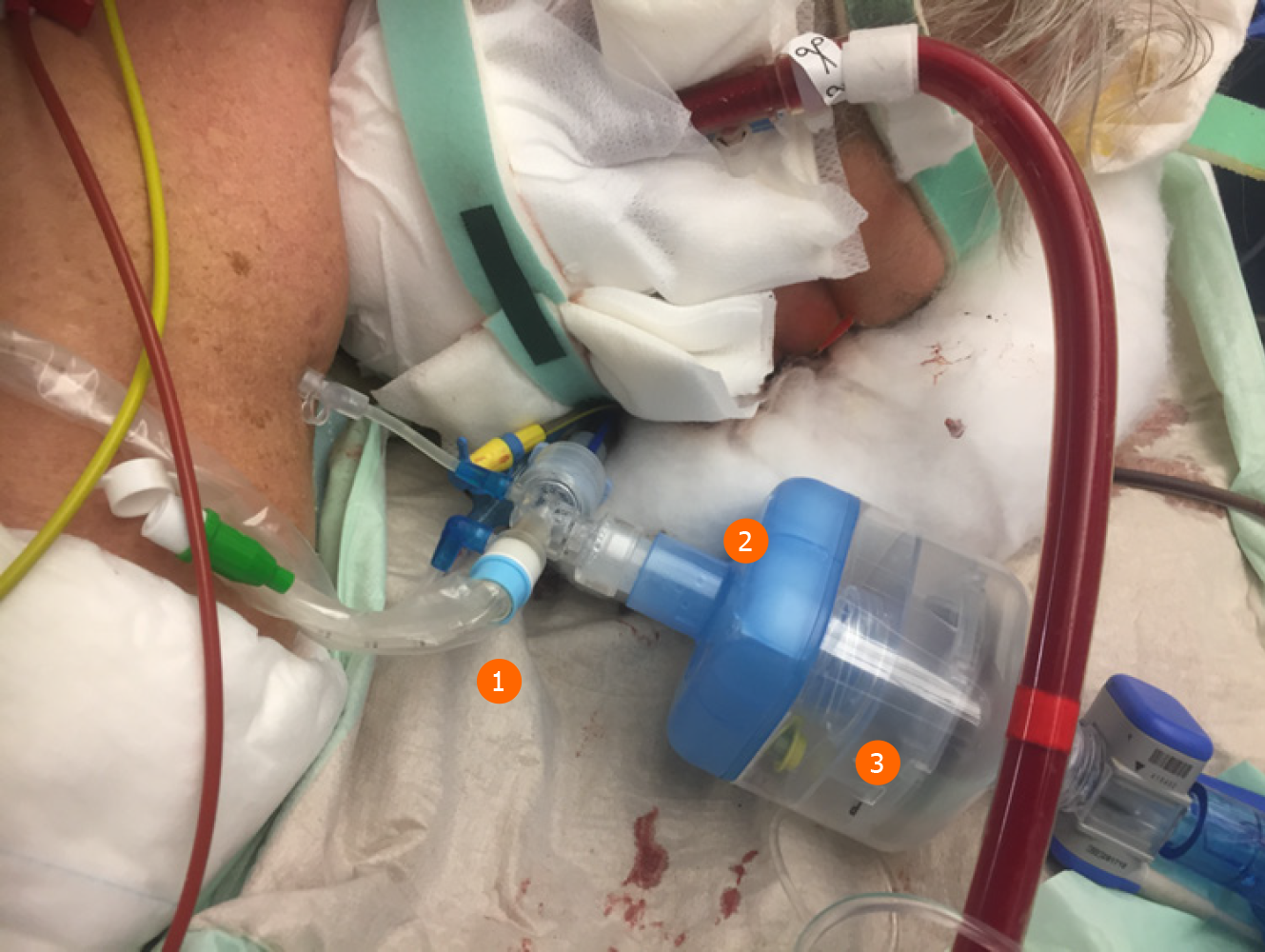
Figure 3 MIRUSTM setup in prone position and veno-venous-extracorporeal membrane oxygenation therapy.
1Closed suction system; 2Bacterial and viral filter and heat and moisture exchanger; 3MIRUSTM reflector.
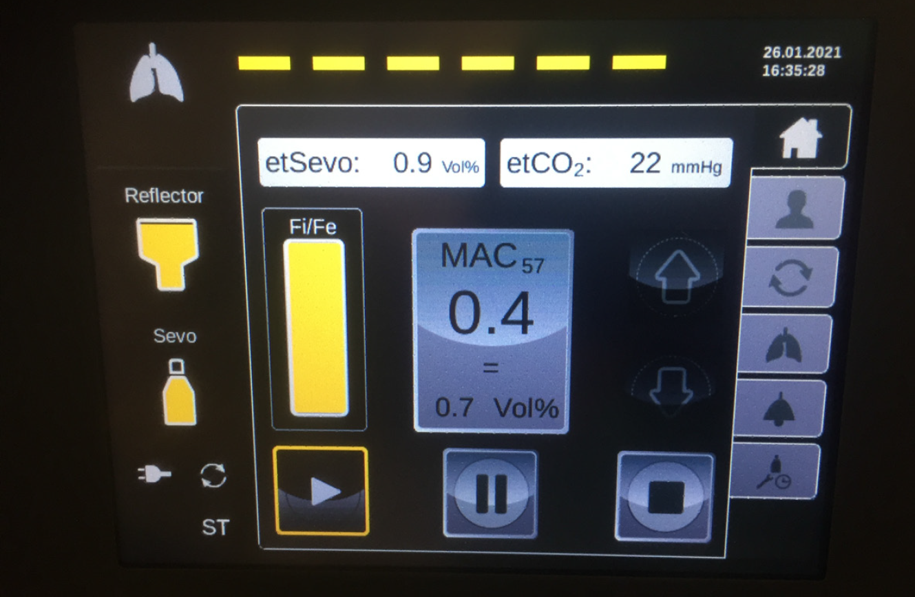
Figure 4 Display of the MIRUSTM sevoflurane controller.
The display shows the setting under normal operation.
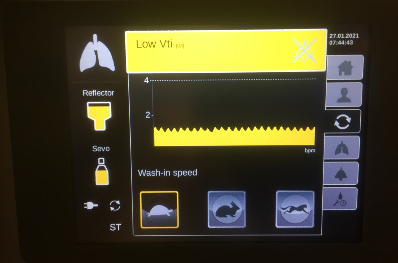
Figure 5 Display of the MIRUSTM controller.
Yellow alarm refers to a low tidal volume. In this case, the wash-in speed “tortoise” should be selected.
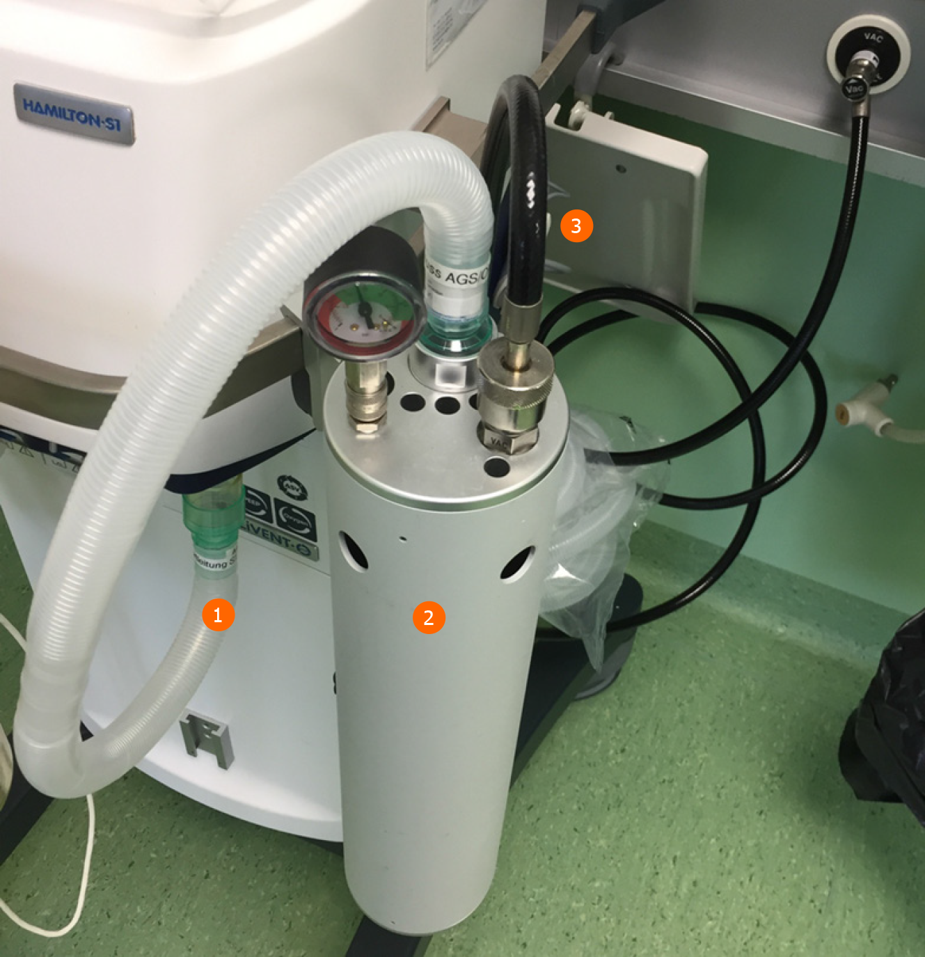
Figure 6 Example of a vacuum-based gas scavenging system (CleanAirTM system).
1Expiration port of the ventilator; 2Open reservoir scavenging system; 3Vacuum line.
- Citation: Bellgardt M, Özcelik D, Breuer-Kaiser AFC, Steinfort C, Breuer TGK, Weber TP, Herzog-Niescery J. Extracorporeal membrane oxygenation and inhaled sedation in coronavirus disease 2019-related acute respiratory distress syndrome. World J Crit Care Med 2021; 10(6): 323-333
- URL: https://www.wjgnet.com/2220-3141/full/v10/i6/323.htm
- DOI: https://dx.doi.org/10.5492/wjccm.v10.i6.323


