©The Author(s) 2025.
World J Clin Pediatr. Dec 9, 2025; 14(4): 108823
Published online Dec 9, 2025. doi: 10.5409/wjcp.v14.i4.108823
Published online Dec 9, 2025. doi: 10.5409/wjcp.v14.i4.108823
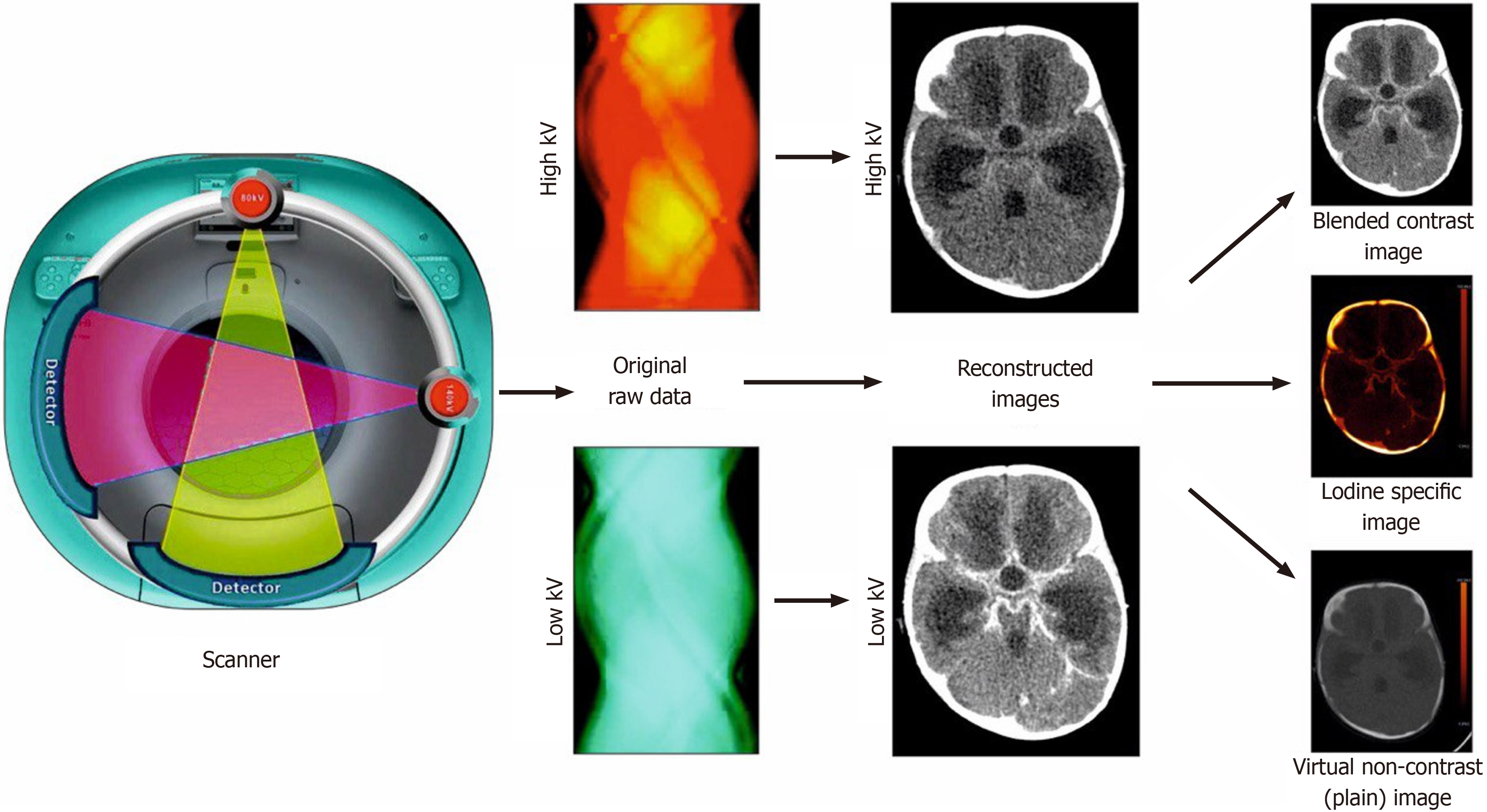
Figure 1 Acquisition of raw data at high and low kV by two different tubes in the scanner.
Reconstruction of images at high and low kV follows. Various applications of dual-energy CT, including the blended contrast-enhanced image, iodine-specific image, and virtual non-contrast (plain) image.
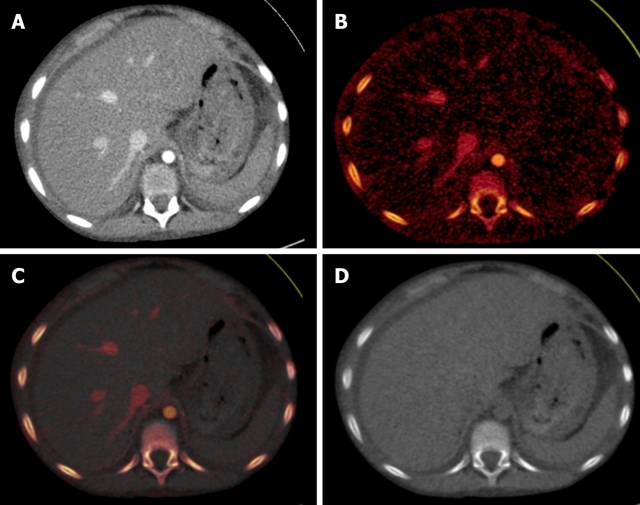
Figure 2 Different reconstructions in a dual-energy CT study.
Axial sections of the upper abdomen at the level of the liver showed normal liver. A: Blended contrast-enhanced image; B: Iodine-specific image; C: Iodine overlay image with superimposed iodine map on gray–scale virtual non-contrast image; D: Virtual non-contrast image.
Figure 3 An 11-year-old female with generalized tonic-clonic seizure and post-ictal drowsiness.
A: Axial section of contrast-enhanced CT head image at 70 kV showed better contrast enhancement of the wall in the ring-enhancing lesion with perilesional edema; B: There was only subtle contrast enhancement in the image obtained at 150 kV.
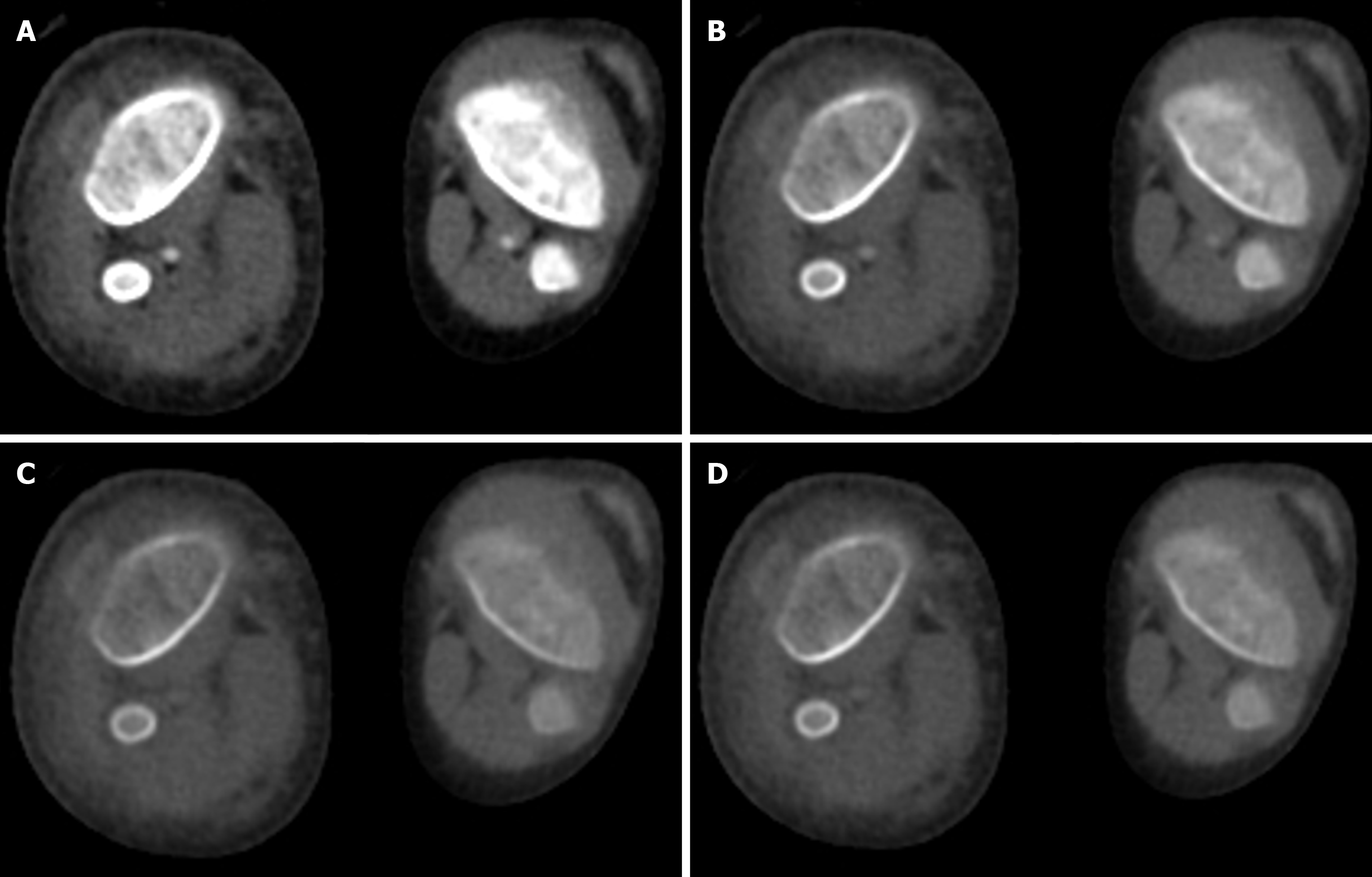
Figure 4 A 4-year-old male with trauma.
Axial virtual monoenergetic images of CT angiography of the bilateral lower limb vessels showed popliteal arteries. A: 40 kV; B: 80 kV; C: 120 kV; D: 160 kV. There was increased conspicuity of the vessels at low kV as compared with high kV. This application allows for the generation of acceptable image quality even with a suboptimal dose of contrast.
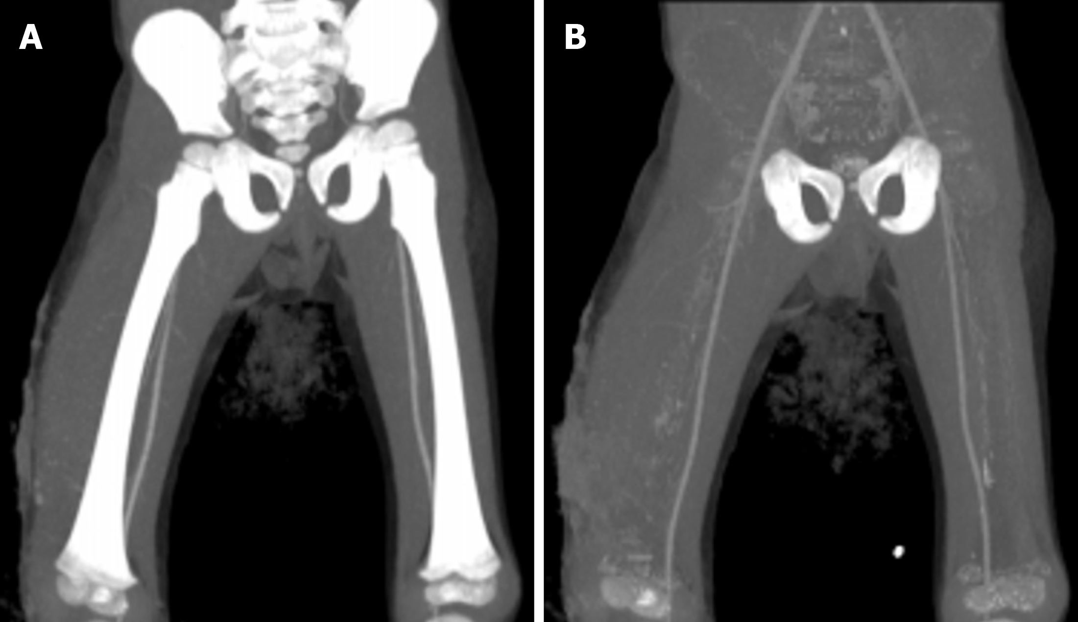
Figure 5 A 4-year-old male with trauma.
Coronal images of CT angiography of bilateral lower limb vessels to show the automated bone removal application of dual energy CT. A: Coronal maximum-intensity projection image showed bilateral superficial femoral arteries overlapped by bones, along with soft tissue swelling along the right thigh; B: Application of the bone removal tool of dual-energy CT enabled better delineation of the arteries and confirmed the absence of any pseudoaneurysm.
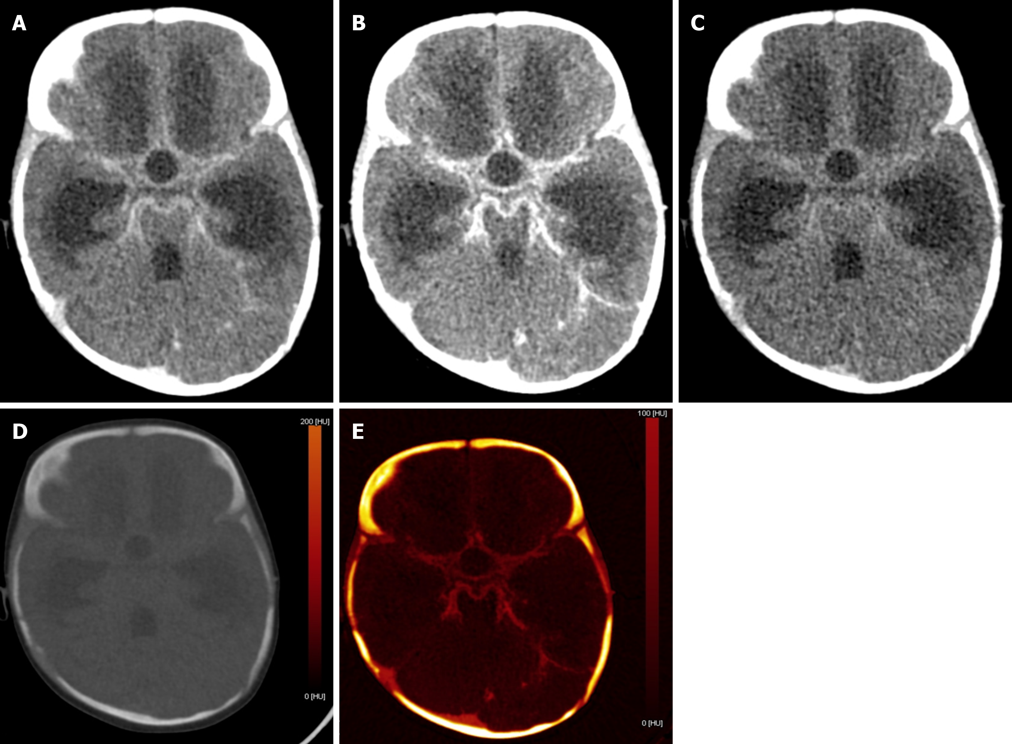
Figure 6 A 7-month-old female with fever and abnormal body movements, suspected of having either a bleed or meningoencephalitis.
A: Axial blended contrast enhanced image showed abnormal leptomeningeal enhancement; B: Image at 70 kV showed marked increased contrast with increased conspicuity of the abnormal enhancement; C: Image at 150 kV showed decreased noise but at the cost of poor contrast; D: Virtual non-contrast image ruled out any abnormal bleed; E: Iodine-specific image showed iodine uptake in the region of hyperdensity, adding to the confidence in the diagnosis of meningitis.
Figure 7 A 4-year-old male with hemophilia A who presented with acute headache and neck rigidity.
A: Axial mixed-enhanced CT head images showed subtle extra-axial high-density lesions (indicated by arrows) in the left frontal and parietal lobes, resulting in a slight midline shift to the opposite side; B: The virtual non-contrast image confirmed that the hyperdensity (arrows) represented an acute bleed and not abnormal enhancement; C: Bone removal tool of dual energy demonstrated the bleed more clearly (arrows); D: Iodine overlay image did not show any iodine uptake in the region of the bleed.
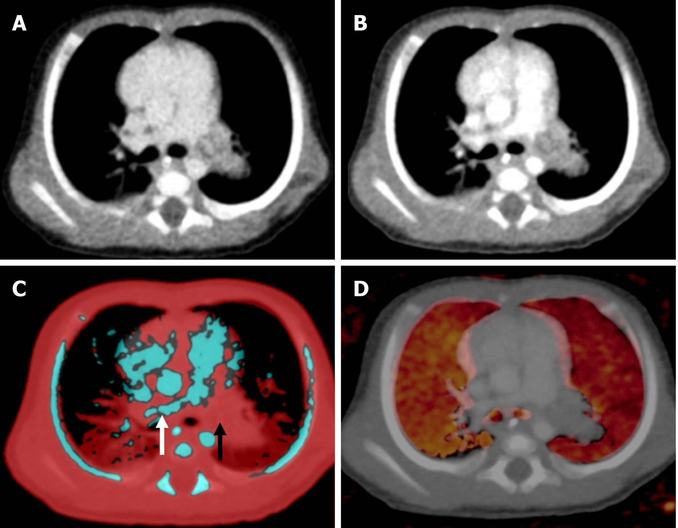
Figure 8 Axial sections of CT pulmonary angiography in a 2-month-old female with infective endocarditis.
A: Complete occlusion of the left pulmonary artery by a thrombus on blended contrast image; B: Complete occlusion of the left pulmonary artery by a thrombus on low kV (80 kV) image, showing increased contrast in the vasculature; C: Lung vessel image showed blue color-coded right pulmonary artery (white arrow) with thrombus being color-coded red in the left pulmonary artery (black arrow), suggesting diminished iodine content; D: Lung blood volume image showed diffusely reduced perfusion in the left lung as compared with the right lung.
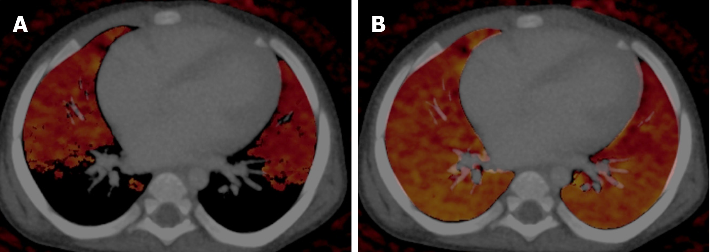
Figure 9 Pitfall related to the incorrect CT threshold used for the assessment of lung blood volume.
A: Iodine defects were seen when the maximum threshold was set at -600 Hounsfield units; B: Lung parenchyma showed normal perfusion when the threshold was changed to -300 Hounsfield units. A higher threshold was needed in children to compensate for the higher lung density.
Figure 10 A case of congenital pulmonary airway malformation in a 5-day-old male.
A: Axial lung window image showed multiple variable-sized cystic lesions interspersed in the right lung; B: Perfused blood volume image showed perfusion deficit in the affected areas; C: Lung vessel image superimposed on lung volume image showed no abnormal vasculature within or surrounding the lesion.
Figure 11 A 10-year-old female with acute pain in the right iliac fossa.
A: Blended dual-energy contrast-enhanced coronal image showed circum
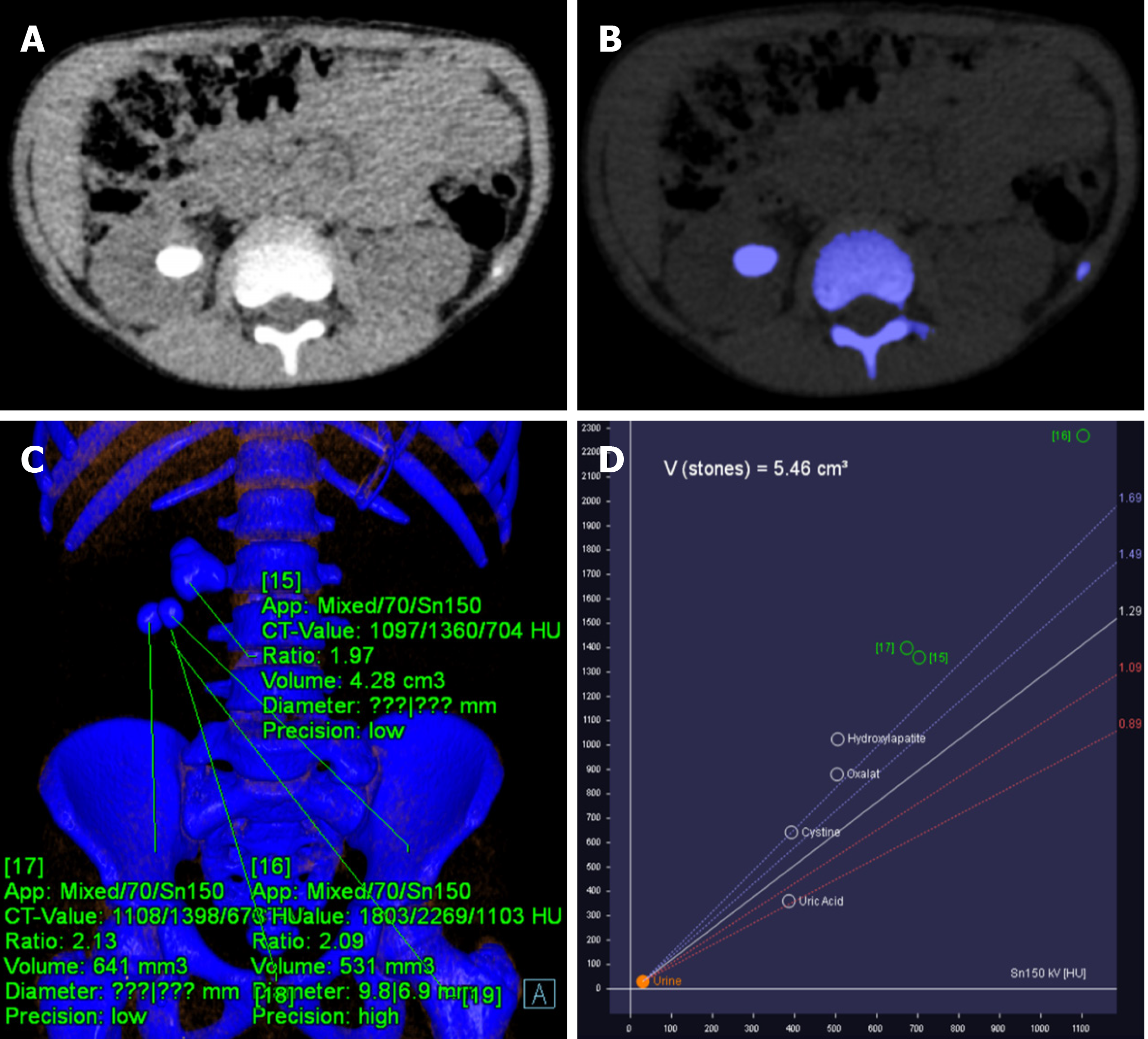
Figure 12 Dual energy scan in nephrolithiasis.
A: Non-contrast CT section of the abdomen showed right renal calculus in the pelvis; B: The calculus was color coded as blue (non-uric acid); C: Attenuation values of calculi on the right side at 70 kV, 150 kV, and blended images; D: The corresponding positions of the attenuation values on the graph indicated non-uric acid composition of the calculi.
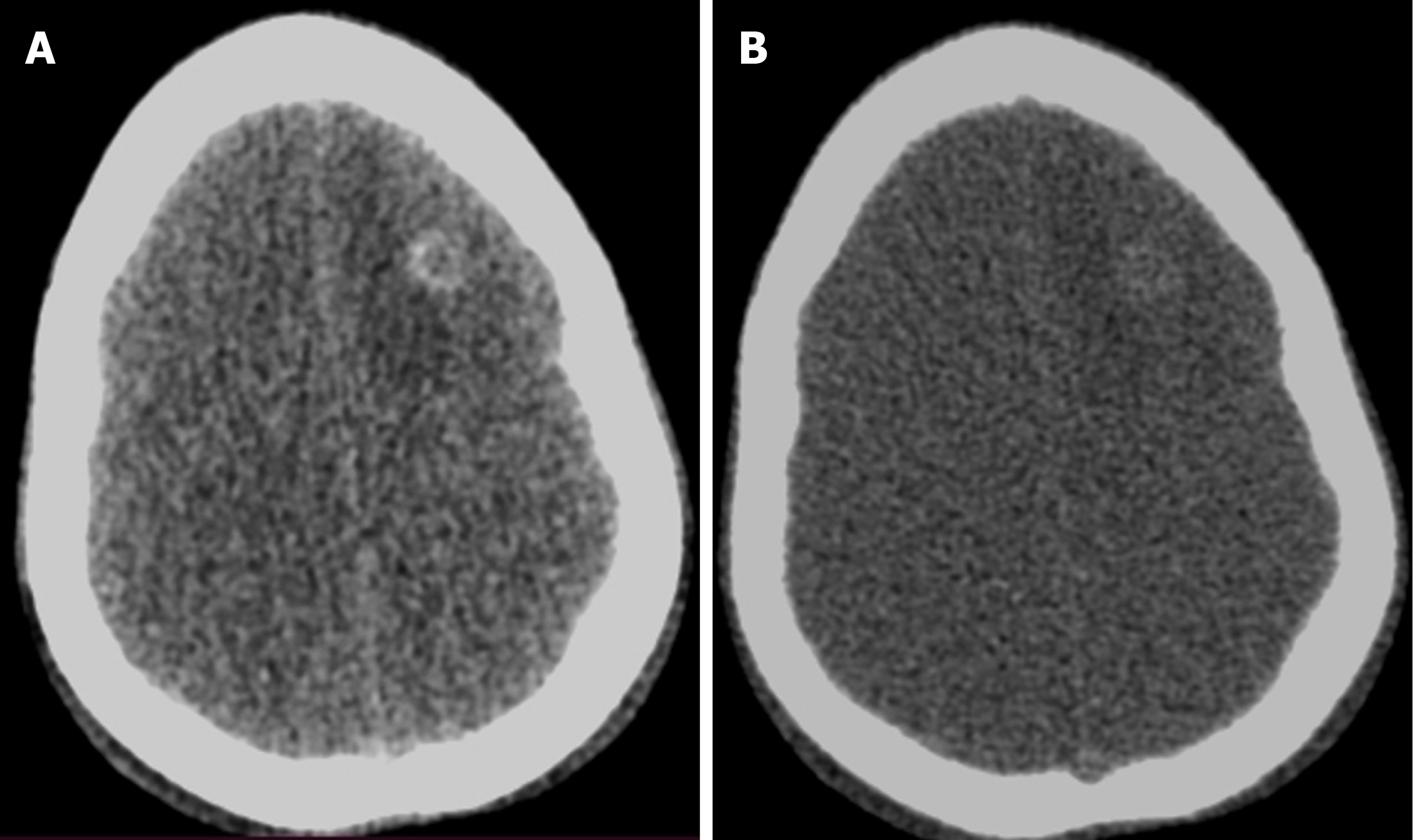
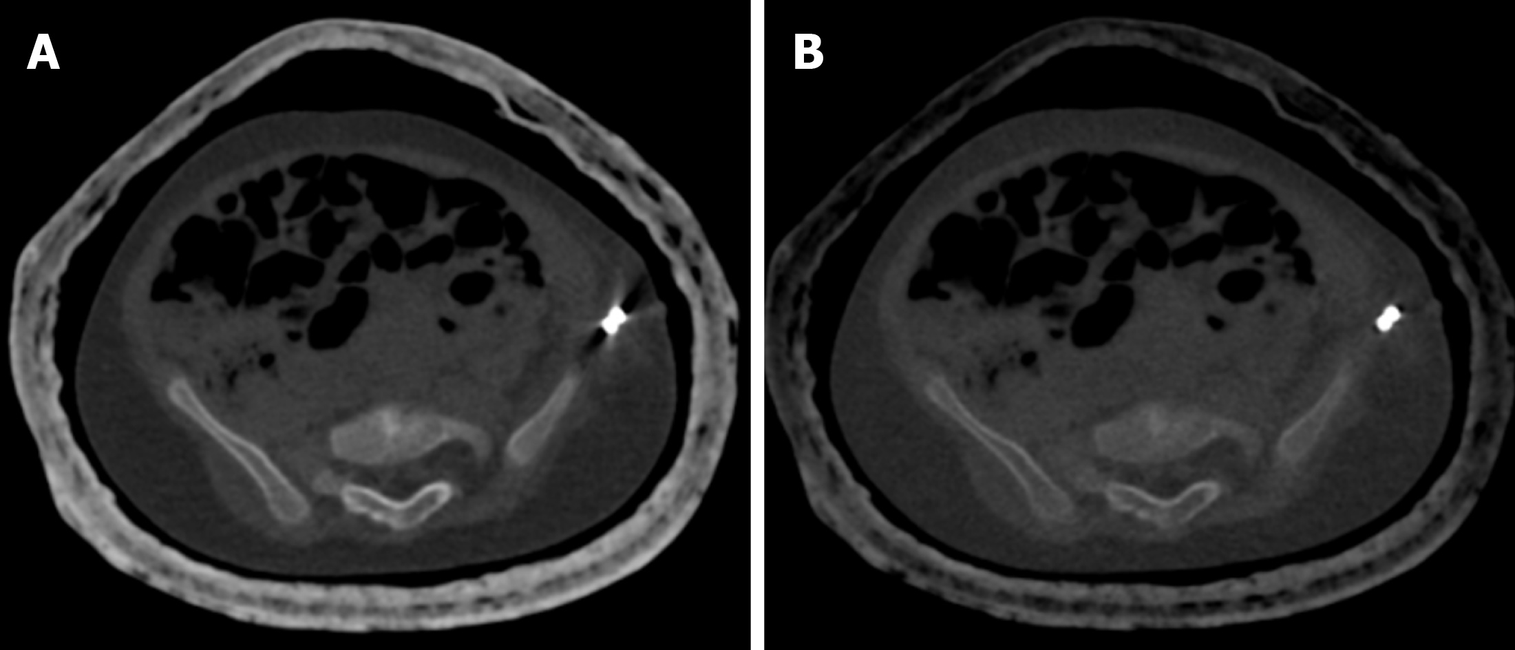
Figure 13 A 2-year-old female who underwent CT of the bilateral hip joints after salter osteotomy for developmental dysplasia of the hip to look for the status of the femur head and acetabulum.
A: Axial bone window image at 70 kV showed marked streak artifacts due to the K-wire inserted along the left iliac bone; B: Axial bone window image at 150 kV showed almost no streak artifacts.
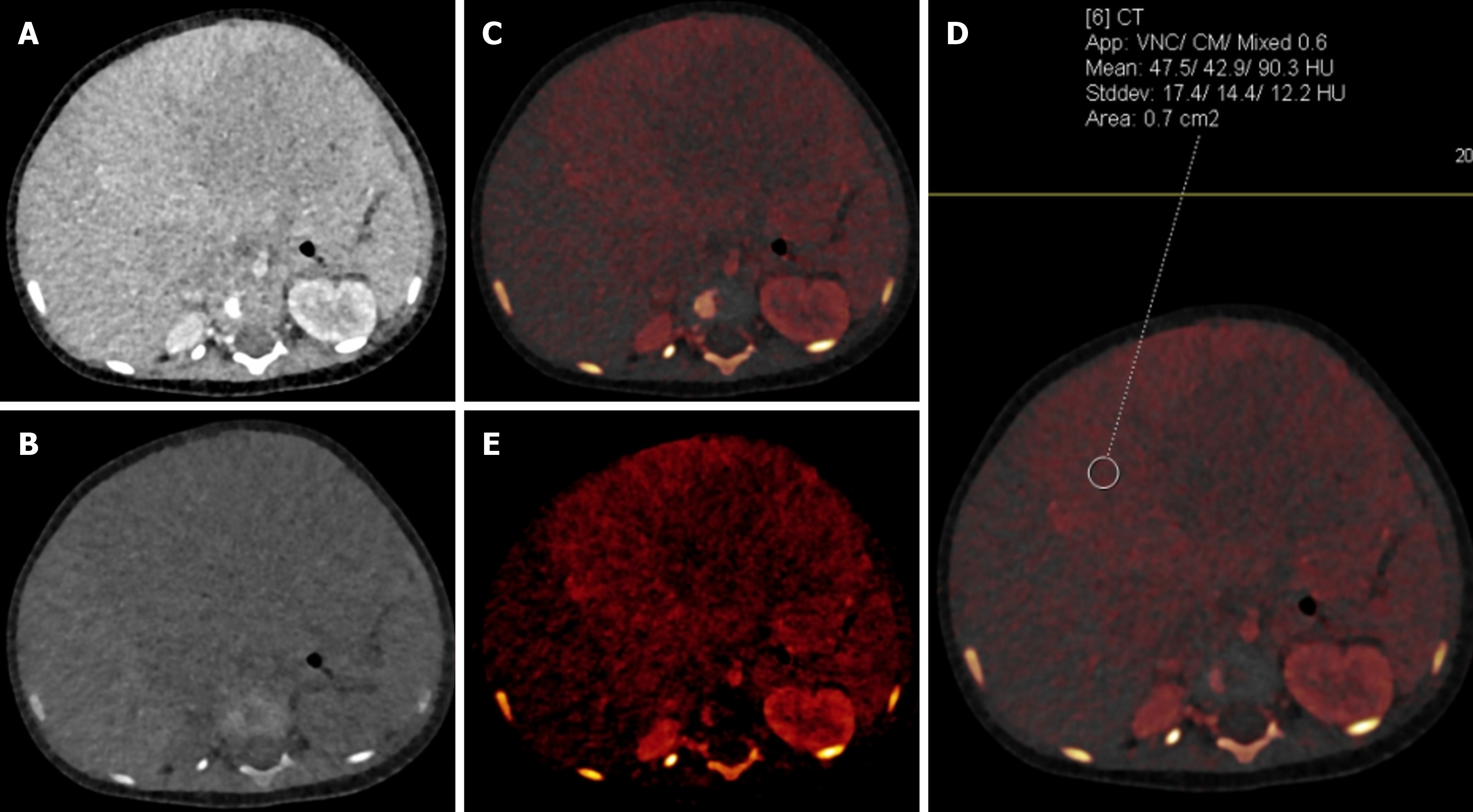
Figure 14 An 11-month-old male with hepatoblastoma.
A: Blended contrast-enhanced axial image showed a large isodense mass in the liver with poor demarcation of the mass with the adjacent liver due to minimal enhancement; B: Virtual non-contrast image showed no evidence of calcification/fat/hemorrhage within the lesion; C and D: Iodine overlay images showed iodine uptake in the periphery of the mass with attenuation on the virtual non-contrast image being 475 Hounsfield units and on the blended image being 903 Hounsfield units, suggesting significant enhancement that was not well appreciated on the blended image; E: Iodine-specific image showed the iodine content in the periphery of the mass to a better advantage.
- Citation: Saini S, Bhatia A, Bansal A, Saxena AK, Sodhi KS. Dual-energy computed tomography in children: Technique and clinical applications. World J Clin Pediatr 2025; 14(4): 108823
- URL: https://www.wjgnet.com/2219-2808/full/v14/i4/108823.htm
- DOI: https://dx.doi.org/10.5409/wjcp.v14.i4.108823


