©The Author(s) 2025.
World J Orthop. Nov 18, 2025; 16(11): 111052
Published online Nov 18, 2025. doi: 10.5312/wjo.v16.i11.111052
Published online Nov 18, 2025. doi: 10.5312/wjo.v16.i11.111052
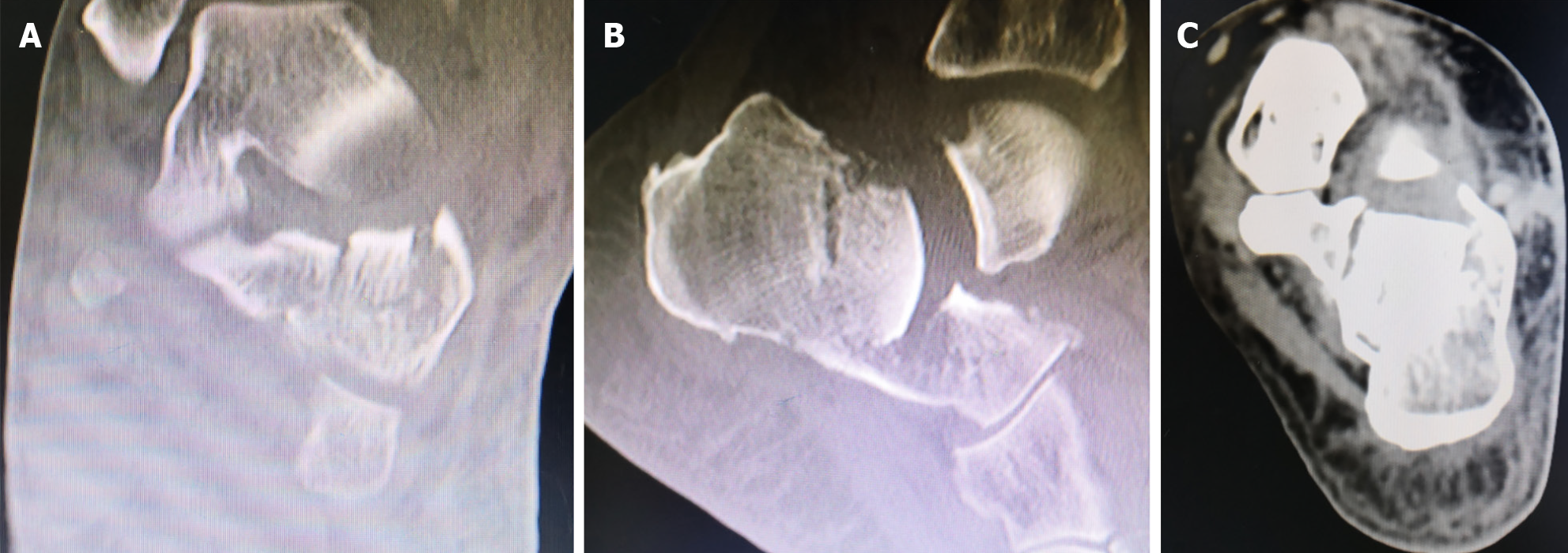
Figure 1 Radiographs revealed the patient’s comminuted calcaneal fracture.
A and B: Anteroposterior (A) and lateral (B) views of the ankle; C: Axial view of the calcaneus; D: Böhler angle was 0°.
Figure 2 Preoperative computed tomography scan findings.
A: Sanders type IV calcaneal fracture; B: Böhler angle was 0°; C: Mild calcaneal widening.
Figure 3 Step 1.
A: The preoperative plan specified a 40° prying angle with the entry point at the anterior margin of the calcaneal tuberosity. The tip of the Steinmann pin was positioned at the deepest point of the depressed fracture fragment; B: Two 2.0 mm Kirschner wires were placed superficially to simulate the intended trajectory and depth of insertion; C: Two 3.0 mm Steinmann pins were then inserted into the calcaneus with approximately 1.5 cm spacing between them, following the trajectory established by the superficial Kirschner wires; D: Fluoroscopic imaging confirmed satisfactory positioning of the superficial Kirschner wires.
Figure 4 Intraoperative fluoroscopy demonstrating the reduction procedure and outcomes.
A: After prying reduction restored the normal Böhler’s angle, the Steinmann pin was used to engage the distal fracture fragment posterior-inferior to the calcaneal body for eversion reduction; B and C: With compression applied to both sides of the calcaneus, fluoroscopy confirmed satisfactory reduction. A 2.5 mm guide wire was then drilled from the posterior calcaneus in an anterior direction; D: Post-reduction photograph.
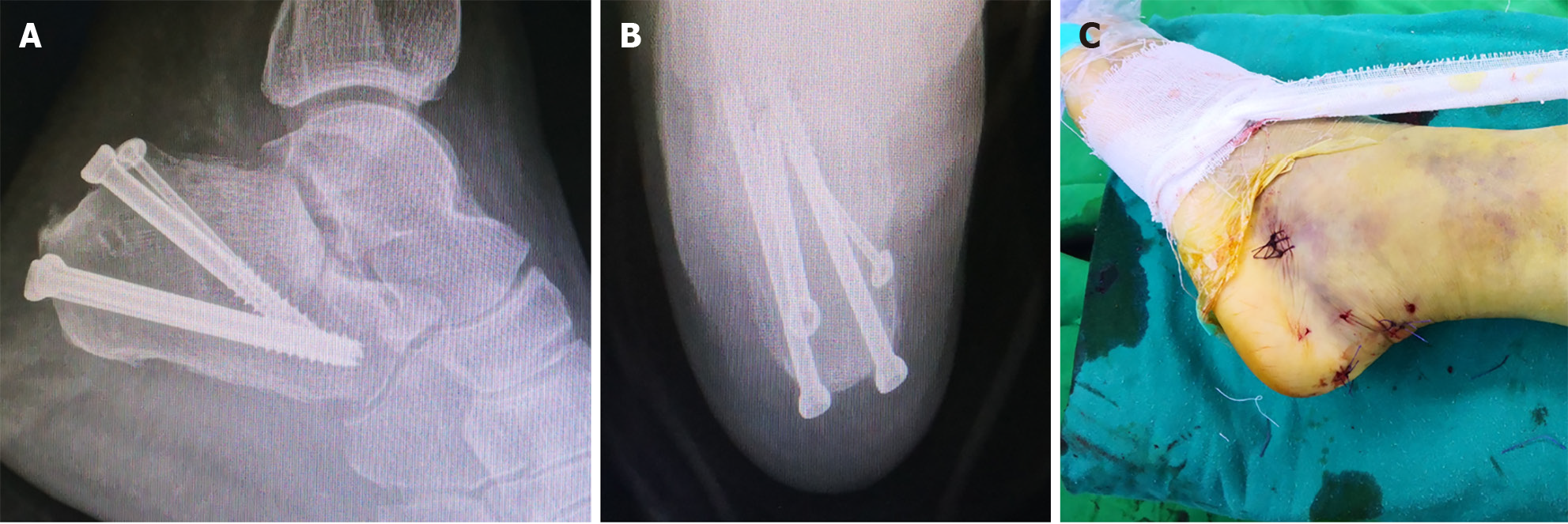
Figure 5 Surgical incision and intraoperative fixation.
A and B: Fluoroscopic lateral (A) and axial (B) views demonstrating percutaneous screw fixation of the calcaneus; C: Photograph of the minimally invasive surgical incision.
Figure 6 Böhler angle changes after screws removal.
After 7 months postoperatively, screws were removed and radiography assessments were carried out in follow-up. A and B: Böhler angle was at 22° at 7 months; C and D: Despite requiring surgical intervention for an ankle fracture due to trauma, at 39 months postoperatively the calcaneal Böhler angle was preserved at the initial 22° reduction.
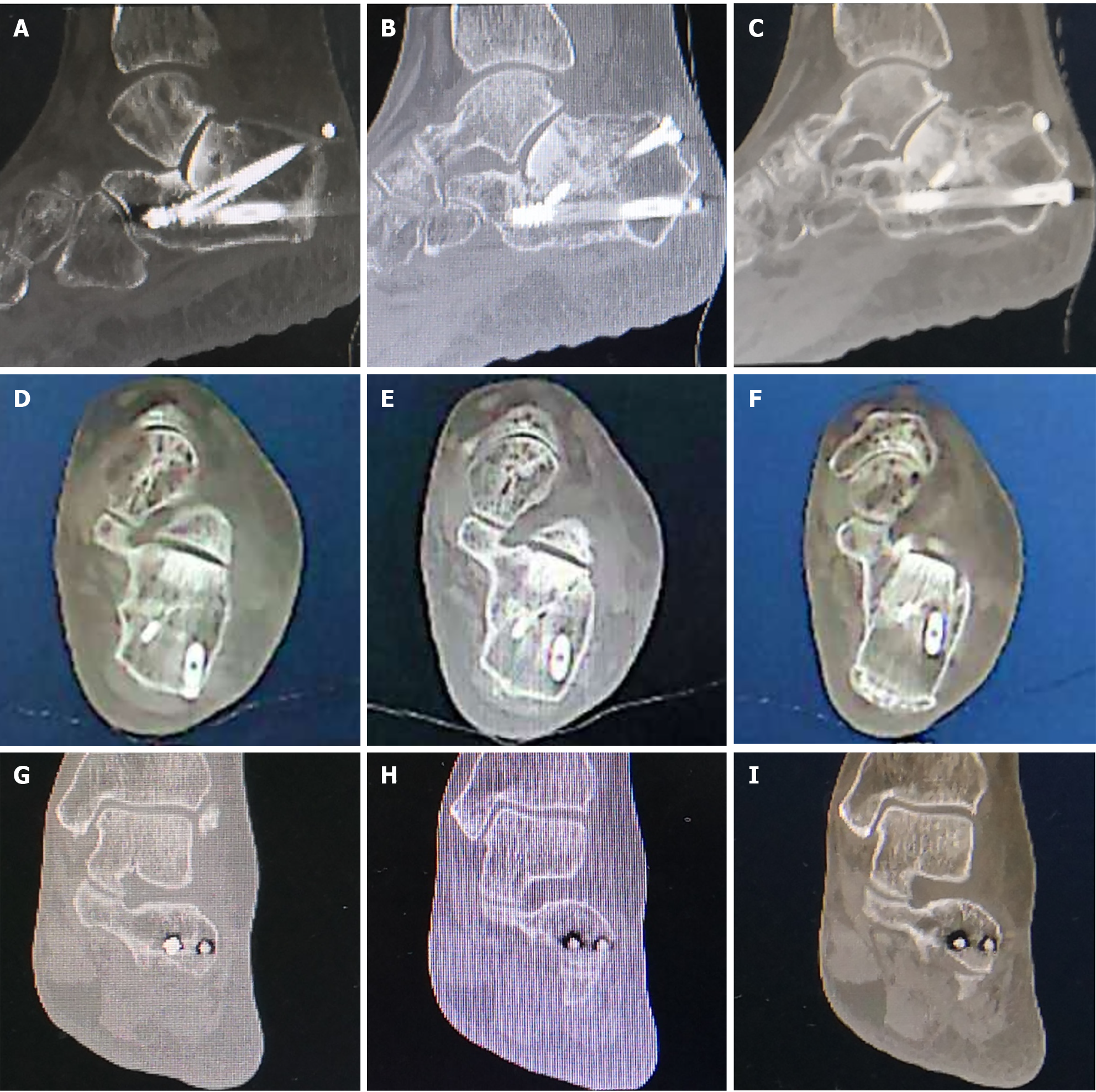
Figure 7 Seven-month postoperative computed tomography scan findings.
A-C: The Böhler’s angle was maintained at 22°; D-F: The calcaneal width was essentially restored to normal width; G-I: The subtalar joint surface demonstrated satisfactory reduction with restored articular congruity.
- Citation: Huang HC, Che YF, Sun H, Xu YS, Gao HY, Tang XS. Traditional Chinese bone-setting combined with percutaneous screw fixation for comminuted calcaneal fractures: A case report and review of literature. World J Orthop 2025; 16(11): 111052
- URL: https://www.wjgnet.com/2218-5836/full/v16/i11/111052.htm
- DOI: https://dx.doi.org/10.5312/wjo.v16.i11.111052


