Copyright
©2014 Baishideng Publishing Group Co.
World J Clin Oncol. May 10, 2014; 5(2): 61-70
Published online May 10, 2014. doi: 10.5306/wjco.v5.i2.61
Published online May 10, 2014. doi: 10.5306/wjco.v5.i2.61
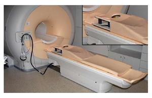
Figure 1 Magnetic resonance imaging scanner with closed bore magnet and a dedicated 8 channel phased-array breast coil (top right).
Technically any magnetic resonance imaging scanner could be used in breast image acquisition. However, in daily practice, field strengths of 1.5 T and 3 T are often used due to higher spatial resolution at similar temporal resolution, providing better diagnostic efficacy.
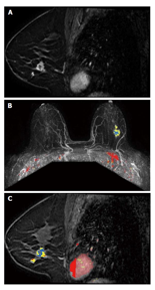
Figure 2 A 48-year-old woman, with positive family history for breast cancer, presented with a palpable lump on the left breast, finally diagnosed as invasive ductolobular carcinoma.
A: Sagittal contrast-enhanced fat-suppressed T1-weighted gradient echo images obtained at 3 T shows a spiculated mass, with rim enhancement and small satellite lesion (multifocal disease); B and C: Color parametric enhancement map in axial postcontrast maximum intensity projection and sagittal projection indicates predominantly a plateau enhancement behavior, with some areas of washout.
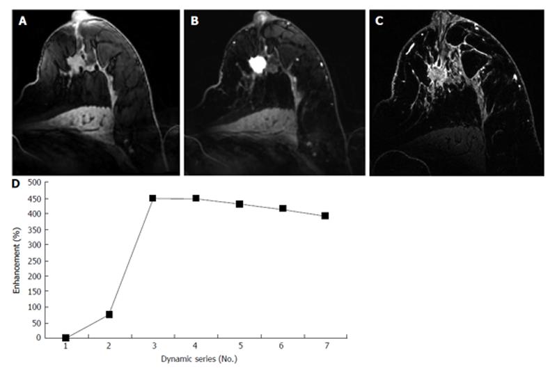
Figure 3 A 62-year-old patient with nipple withdrawal, finally diagnosed as ductolobular carcinoma.
A, B and C: Axial T1-weighted gradient-echo images obtained at 7 T before and after contrast injection. An irregular mass with spiculated margins can be observed on pré-contrast imaging (A). An intense homogeneous enhancement (B) and a rapid wash-out kinetic curve (D) can be observed following contrast administration. In Figure 3C, an ultra-high-resolution T1-weighted gradient-echo sequence with fat suppression was performed, and the morphological aspects of the lesion can be more clearly seen.
- Citation: Menezes GL, Knuttel FM, Stehouwer BL, Pijnappel RM, van den Bosch MA. Magnetic resonance imaging in breast cancer: A literature review and future perspectives. World J Clin Oncol 2014; 5(2): 61-70
- URL: https://www.wjgnet.com/2218-4333/full/v5/i2/61.htm
- DOI: https://dx.doi.org/10.5306/wjco.v5.i2.61


