
















Copyright: ©Author(s) 2026.
World J Clin Oncol. May 24, 2026; 17(5): 119615
Published online May 24, 2026. doi: 10.5306/wjco.v17.i5.119615
Published online May 24, 2026. doi: 10.5306/wjco.v17.i5.119615
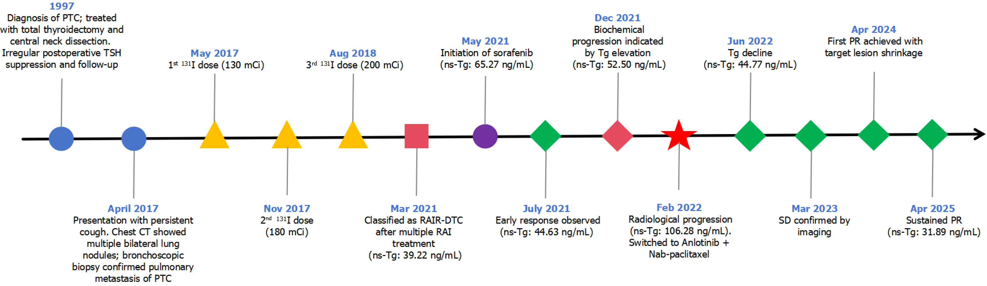
Figure 1 Clinical timeline illustrating the disease trajectory, therapeutic interventions, and dynamic changes in thyroglobulin levels.
The timeline demonstrates the clinical progression from the initial surgical intervention and radioactive iodine treatments to sequential targeted therapies. The red star marks a critical clinical juncture in February 2022, when significant radiological progression prompted the transition to the combined anlotinib and nab-paclitaxel regimen. This strategic intervention ultimately resulted in a sustained partial response and continuous decline in thyroglobulin levels. PTC: Papillary thyroid carcinoma; TSH: Thyroid-stimulating hormone; CT: Computed tomography; RAIR-DTC: Radioiodine-refractory differentiated thyroid cancer; RAI: Radioactive iodine; ns-Tg: Non-stimulated thyroglobulin; SD: Stable disease; PR: Partial response.
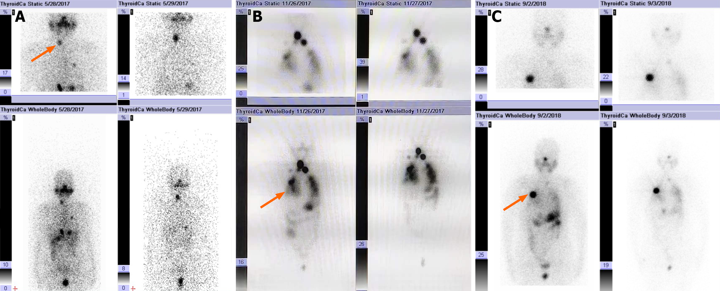
Figure 2 Serial 131I whole-body scintigraphy following three courses of radioiodine therapy.
A: Initial post-therapeutic imaging revealed cervical thyroid remnants and scattered radioactive iodine uptake in both lungs; B: Subsequent scans after the second dose demonstrated persistent neck uptake, along with a more extensive, diffuse distribution in the pulmonary regions; C: By the third cycle, no significant thyroid tissue was visible in the neck, and pulmonary foci had diminished, although an active left subclavian lymph node remained.
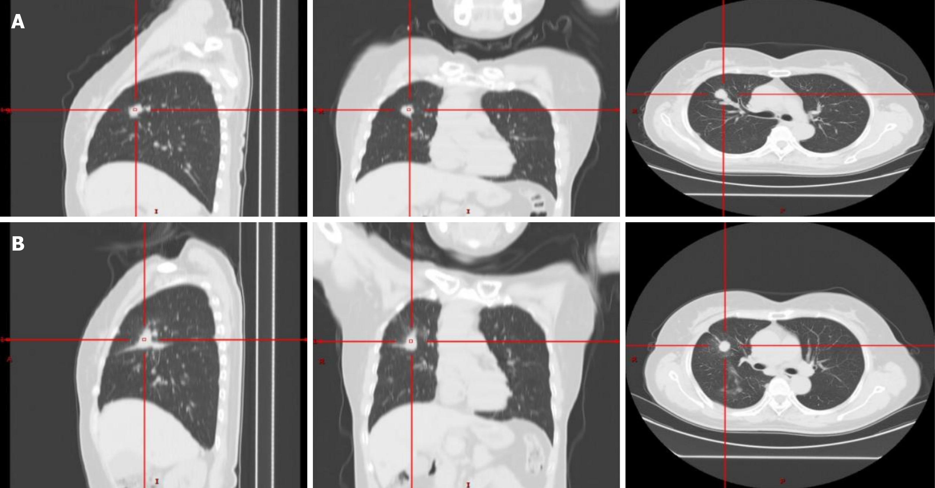
Figure 3 Progressive pulmonary metastases on computed tomography following completion of radioiodine therapy.
A: The computed tomography (CT) scan obtained 6 months after the third 131I treatment shows multiple metastatic nodules in both lungs; B: The CT scan obtained 21 months after the third 131I treatment demonstrates an increase in the size of pulmonary metastases compared with panel A, indicating structural disease progression despite prior radioiodine therapy.
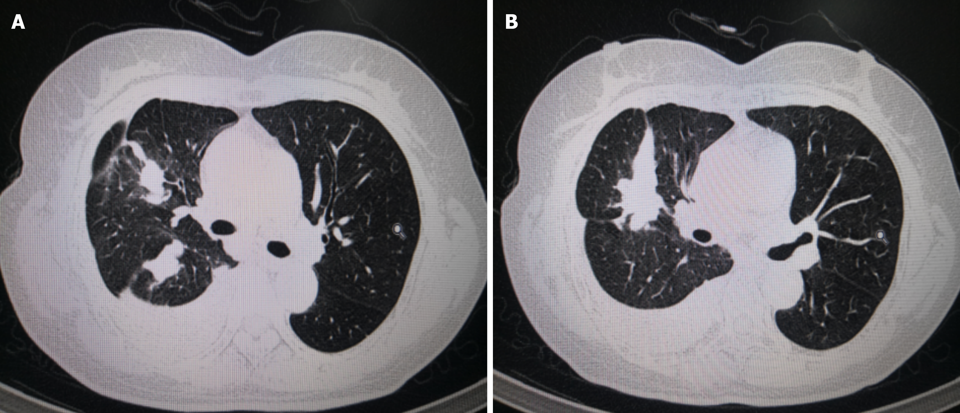
Figure 4 Diffuse pulmonary disease and pleural effusion at the time of radioiodine-refractory differentiated thyroid cancer diagnosis.
A and B: A computed tomography scan performed 33 months after the third 131I treatment reveals widespread bilateral pulmonary metastases. Additionally, obstructive pneumonia within the right upper lobe and a massive right-sided pleural effusion are evident. These findings, along with the diminishing radioiodine uptake on serial 131I scans, support the diagnosis of radioiodine-refractory differentiated thyroid cancer.
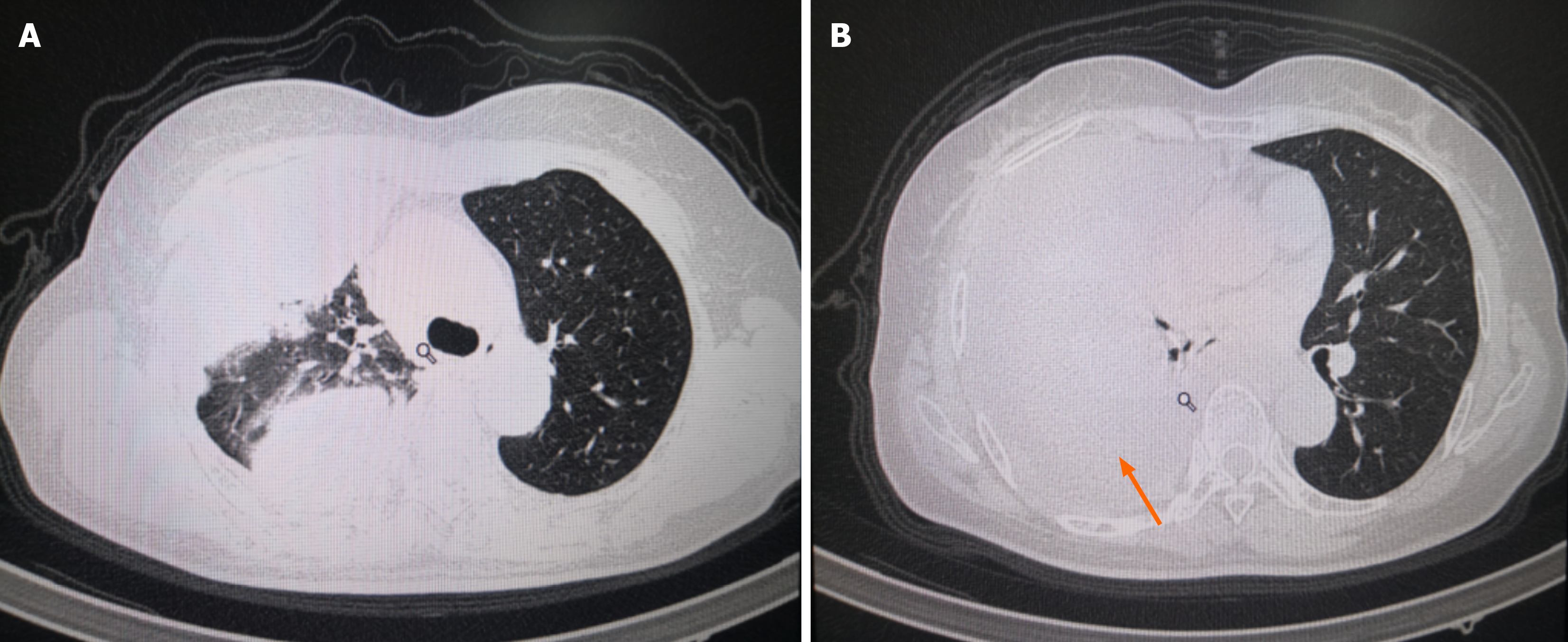
Figure 5 Radiologic progression during sorafenib therapy.
A and B: A computed tomography scan obtained in February 2022, following several months of firstline sorafenib treatment, shows extensive bilateral pulmonary metastases and a substantial increase in the rightsided pleural effusion compared with prior imaging (arrows). These findings, together with rising serum thyroglobulin levels and worsening respiratory symptoms, indicate progressive disease under sorafenib.
- Citation: Zhou CY, Ren XY, Li HH, Pan XM, Lan X, Duan D. Anlotinib plus albumin-bound paclitaxel induction-maintenance for sorafenib-refractory radioiodine-refractory differentiated thyroid cancer: A case report and review of literature. World J Clin Oncol 2026; 17(5): 119615
- URL: https://www.wjgnet.com/2218-4333/full/v17/i5/119615.htm
- DOI: https://dx.doi.org/10.5306/wjco.v17.i5.119615