©The Author(s) 2025.
World J Clin Oncol. Jul 24, 2025; 16(7): 107109
Published online Jul 24, 2025. doi: 10.5306/wjco.v16.i7.107109
Published online Jul 24, 2025. doi: 10.5306/wjco.v16.i7.107109
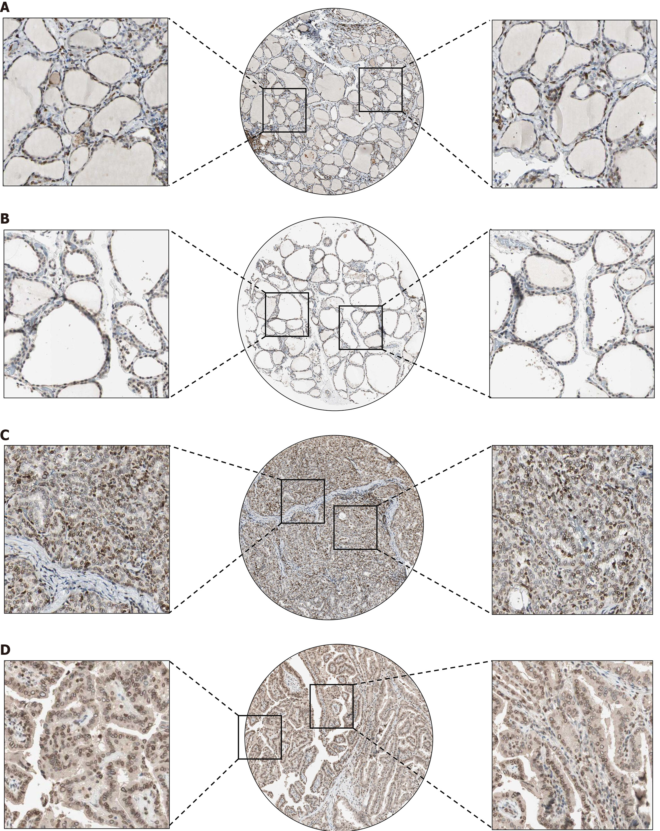
Figure 1 Thymopoietin protein in The Human Protein Atlas showed high expression in papillary thyroid carcinoma.
A and B: Normal thyroid tissue; C and D: Papillary thyroid carcinoma tissue.
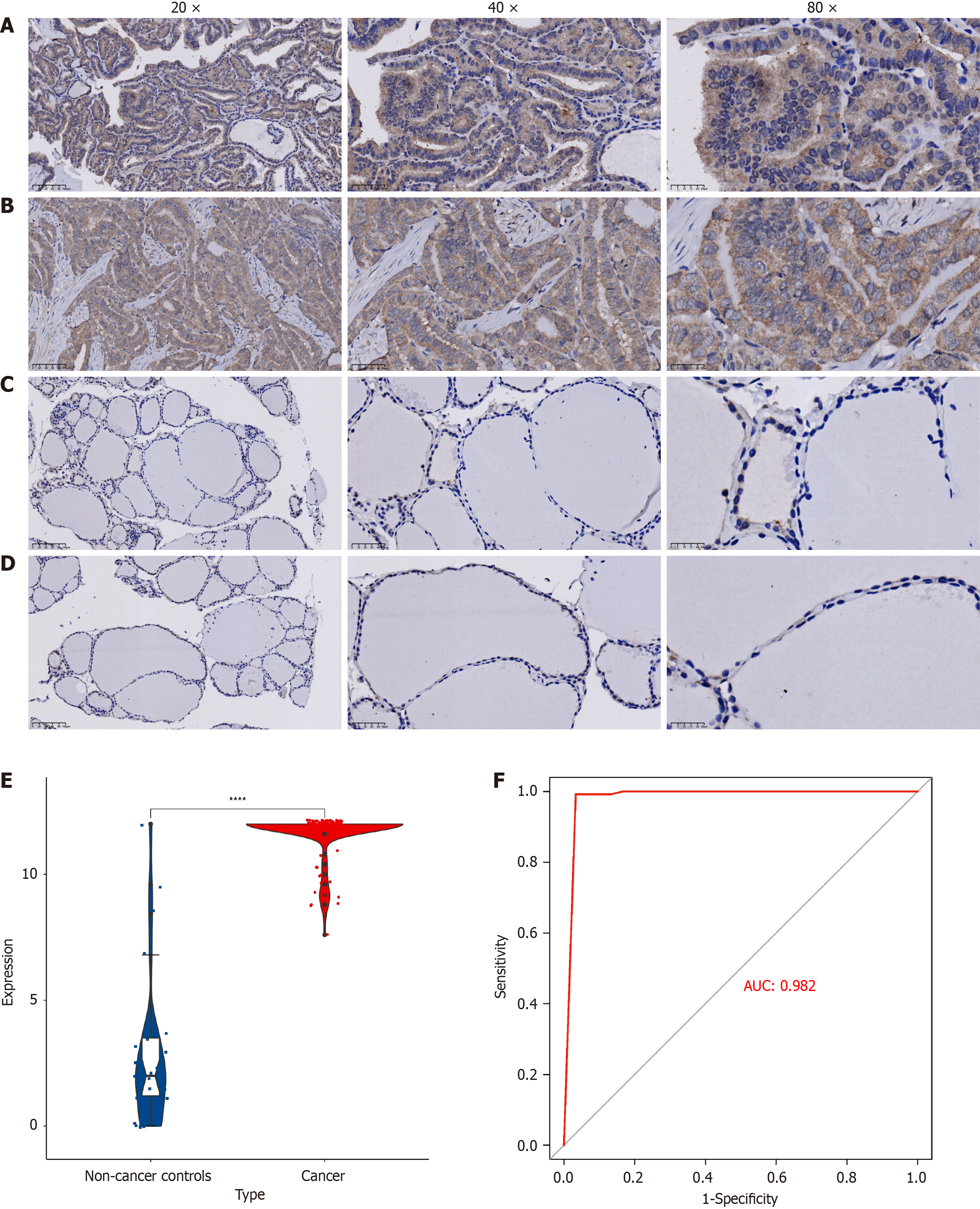
Figure 2 Thymopoietin protein in immunohistochemistry is highly expressed in papillary thyroid carcinoma.
A and B: Thyroid cancer tissue; C and D: Normal thyroid tissue; E: Represents horizontal violin diagram; F: Thymopoietin differential receiver operating characteristic curve between papillary thyroid carcinoma (PTC) and non-PTC.
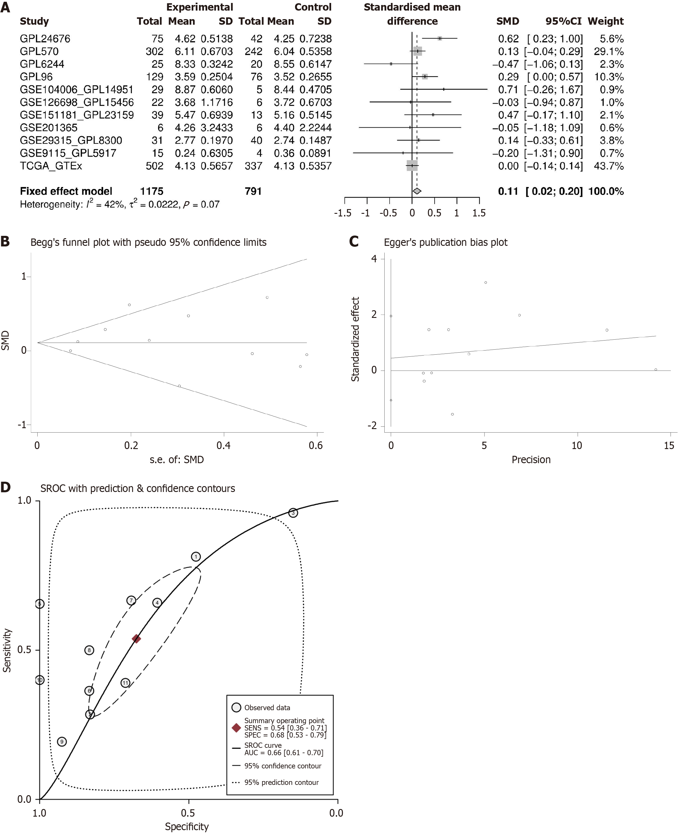
Figure 3 Thymopoietin mRNA exhibits higher expression in papillary thyroid carcinoma.
A: Comparison with normal tissues shows higher thymopoietin (TMPO) mRNA expression in papillary thyroid carcinoma (PTC); B and C: Bias tests reveal no significant publication bias; D: Summary receiver operating characteristic indicates TMPO has discriminative capability in PTC. SMD: Standardized mean difference; SROC: Summary receiver operating characteristic.
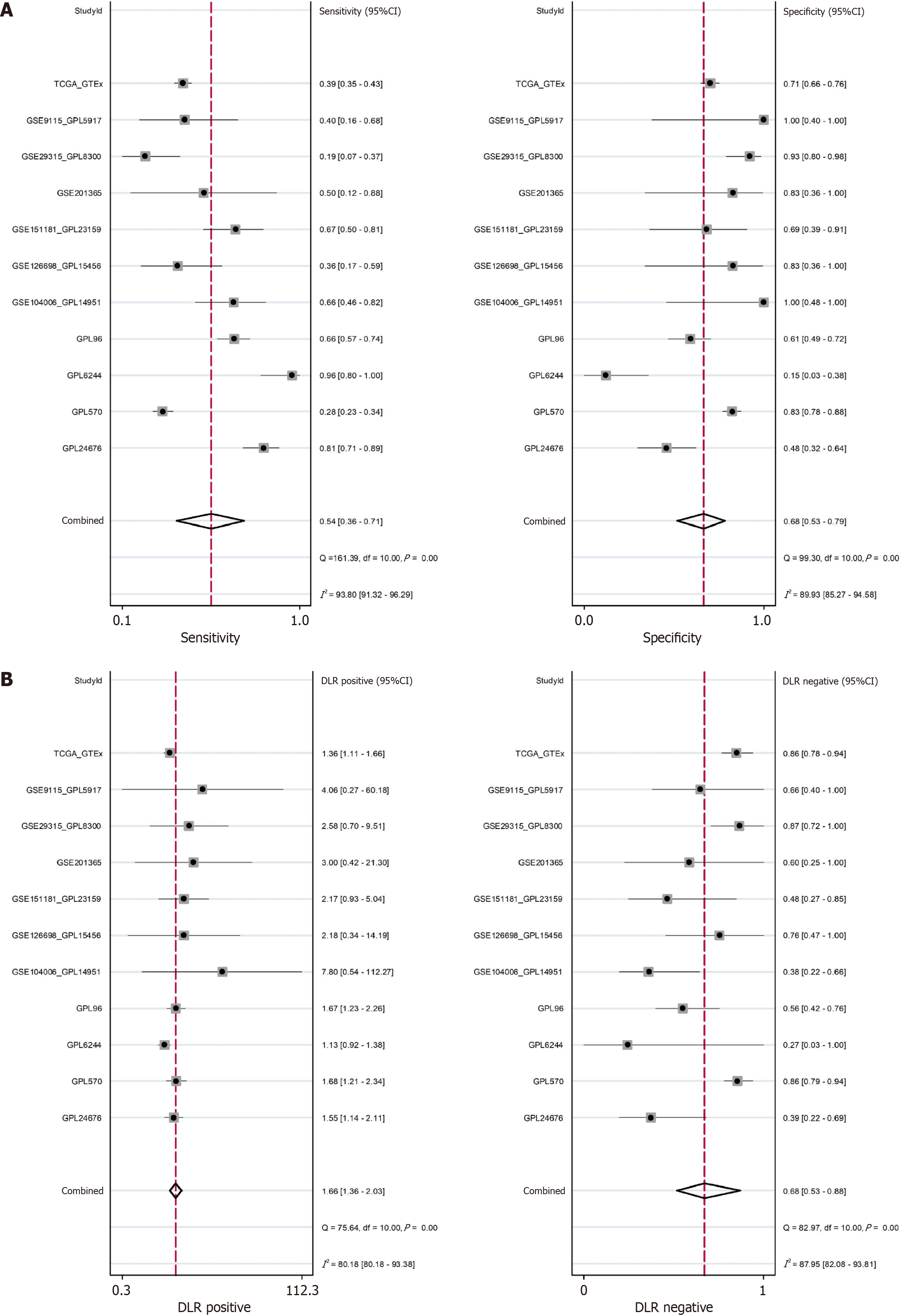
Figure 4 Diagnostic test evaluation of thymopoietin.
A: Sensitivity and specificity; B: Positive likelihood ratio and negative likelihood ratio.
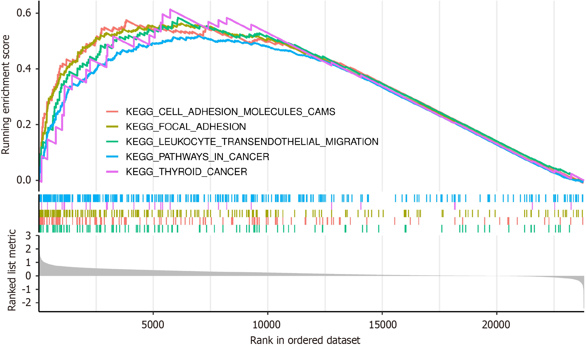
Figure 5
Gene Set Enrichment Analysis results for thymopoietin.
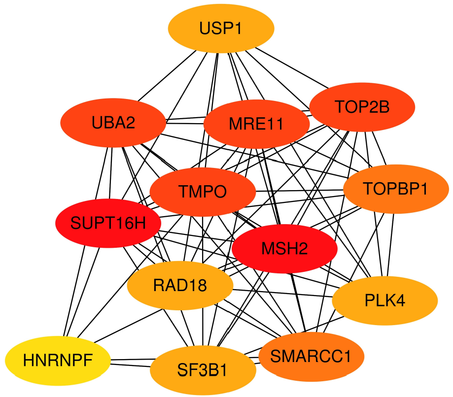
Figure 6
Analysis of thymopoietin protein interaction network.
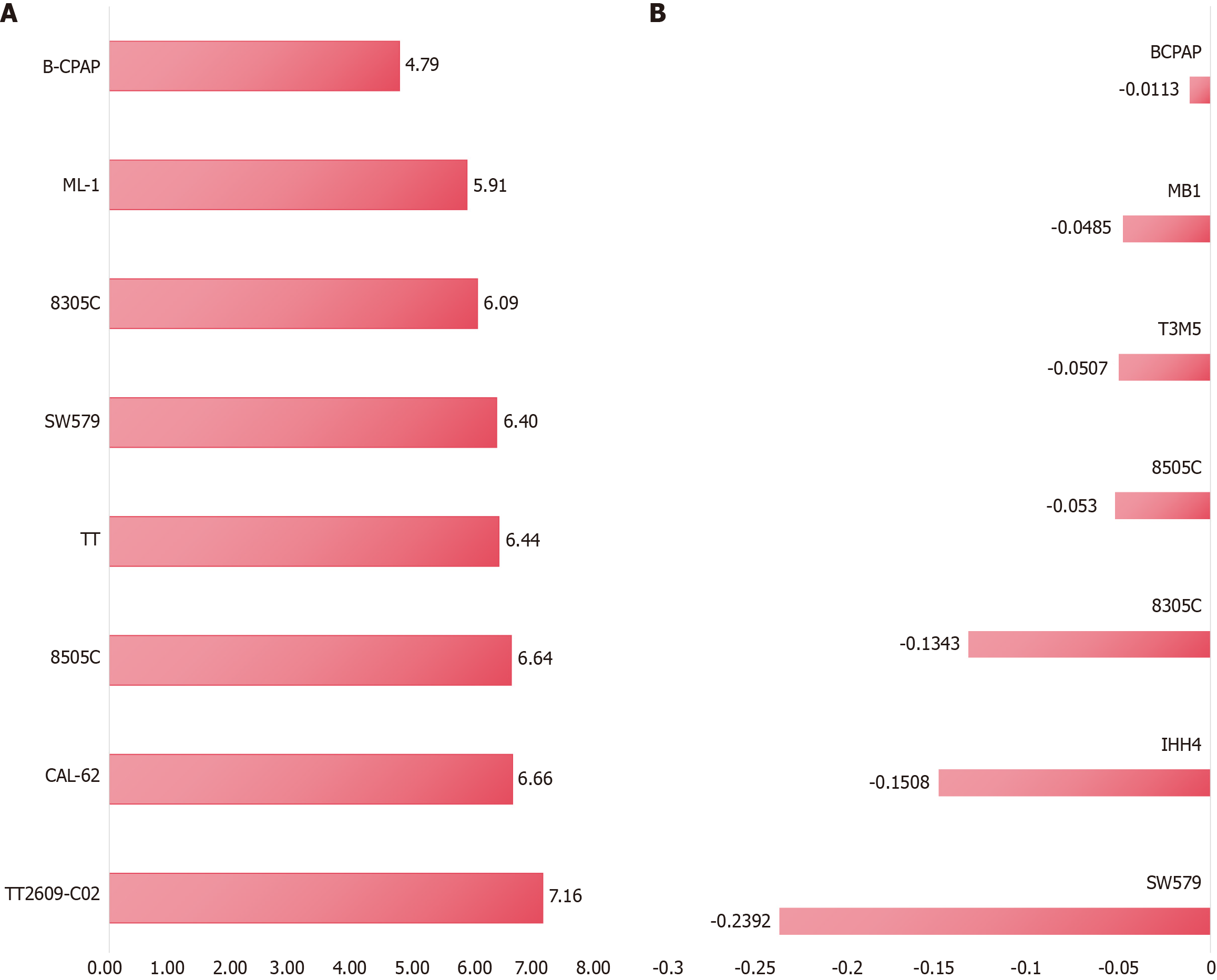
Figure 7 Growth of thymopoietin in papillary thyroid carcinoma cell lines and cell line growth inhibition after knockout of the thymopoietin gene based on Crispr knockout screen technology.
A: Based on Cancer Cell Line Encyclopedia results; B: Based on Crispr knockout screen results.
- Citation: Song C, Pang YY, Lu SY, Li B, Li DM, He RQ, Qin DY, Li SD, Qv N, Chen YM, Chen G, He J, Jiang XB. Molecular mechanisms of thymopoietin in papillary thyroid cancer: Multiplatform gene expression data, gene knockout screening, and in-house immunohistochemistry. World J Clin Oncol 2025; 16(7): 107109
- URL: https://www.wjgnet.com/2218-4333/full/v16/i7/107109.htm
- DOI: https://dx.doi.org/10.5306/wjco.v16.i7.107109


