©The Author(s) 2025.
World J Clin Oncol. May 24, 2025; 16(5): 105322
Published online May 24, 2025. doi: 10.5306/wjco.v16.i5.105322
Published online May 24, 2025. doi: 10.5306/wjco.v16.i5.105322
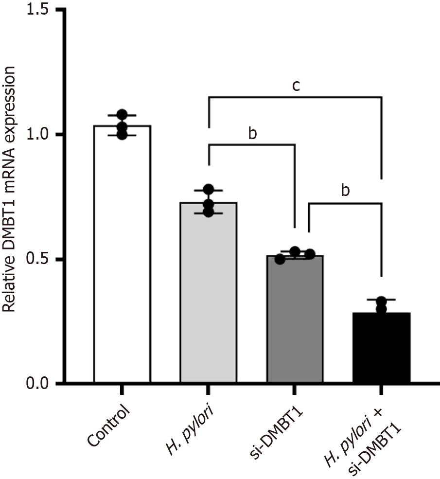
Figure 1 Quantitative real-time polymerase chain reaction to detect the relative expression of deleted in malignant brain tumors 1 mRNA in AGS cells of each group.
bP < 0.001, cP < 0.0001. H. pylori: Helicobacter pylori; DMBT1: Deleted in malignant brain tumors 1.
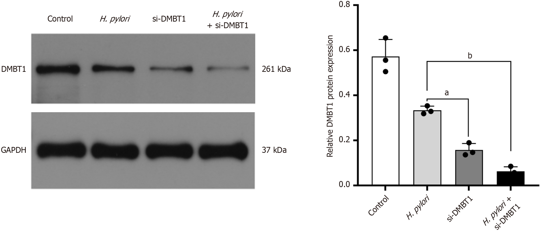
Figure 2 Western blot detection of the relative expression of deleted in malignant brain tumors 1 mRNA in AGS cells of each group.
aP < 0.01, bP < 0.001. H. pylori: Helicobacter pylori; DMBT1: Deleted in malignant brain tumors 1; GAPDH: Glyceraldehyde-3-phosphate dehydrogenase.
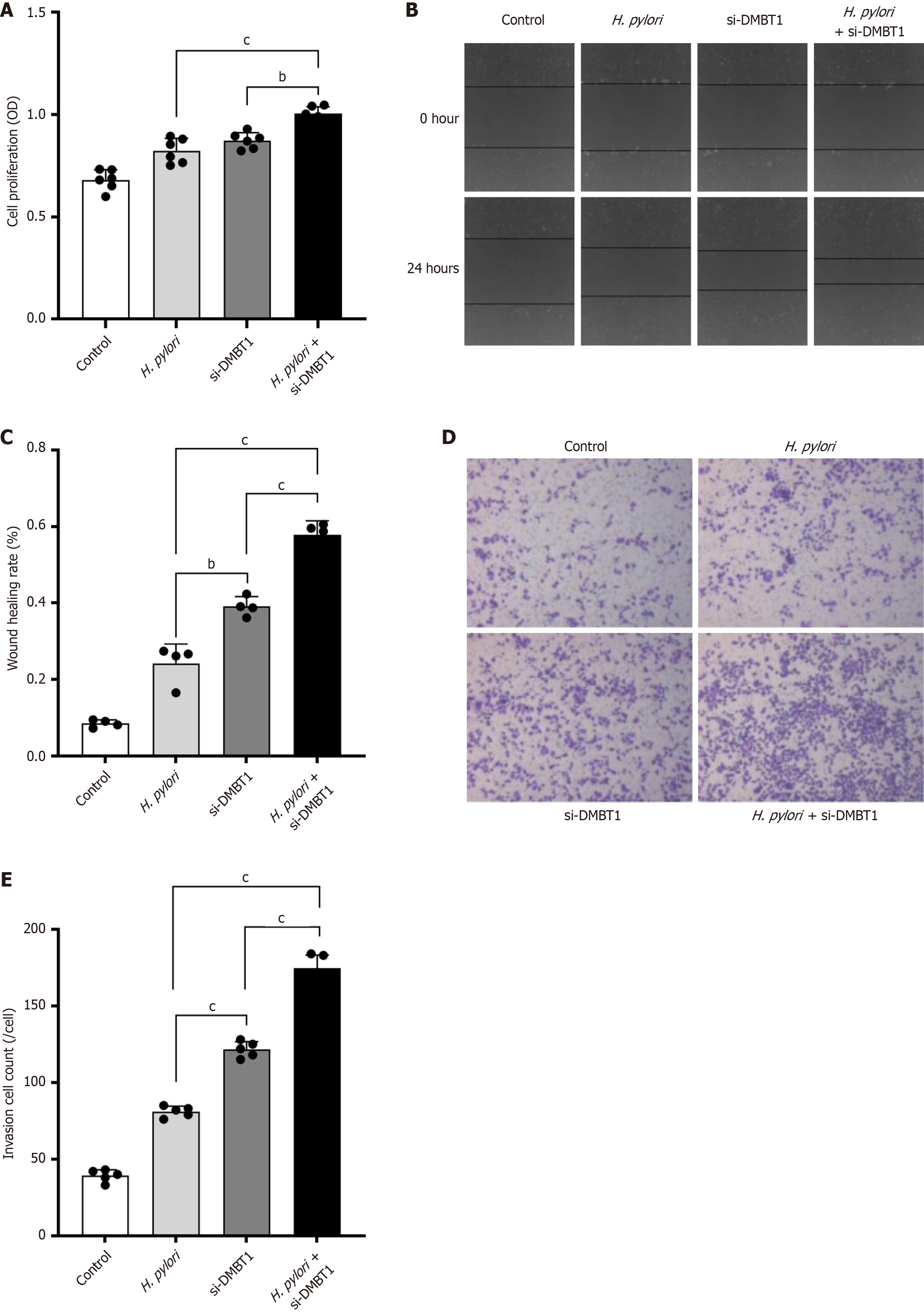
Figure 3 Cell counting kit-8, scratch, and Transwell assays to detect the biological behavior in AGS cells of each group.
A: Cell counting kit-8 assay was employed to detect the proliferation ability of AGS cells in each group; B and C: Cell scratch assay to detect the migration ability of AGS cells in each group (40 ×); D and E: Transwell assay was performed in AGS cells to detect the invasion ability in each group (40 ×). bP < 0.001,cP < 0.0001. H. pylori: Helicobacter pylori; DMBT1: Deleted in malignant brain tumors 1; OD: Optical density.
- Citation: Zhou X, Wang LQ, Song S, Xu M, Li CP. Helicobacter pylori infection promotes the progression of gastric cancer by regulating the expression of DMBT1. World J Clin Oncol 2025; 16(5): 105322
- URL: https://www.wjgnet.com/2218-4333/full/v16/i5/105322.htm
- DOI: https://dx.doi.org/10.5306/wjco.v16.i5.105322


