©The Author(s) 2025.
World J Clin Oncol. Jan 24, 2025; 16(1): 96131
Published online Jan 24, 2025. doi: 10.5306/wjco.v16.i1.96131
Published online Jan 24, 2025. doi: 10.5306/wjco.v16.i1.96131
Figure 1 Hematoxylin-eosin staining.
A: Showed CK (-), CD38 (+), CD138 (+), EMA (+), CyclinD1 (-), k (-), λ (+), CD20 (-), CD68 (-), Ki-67 (5%+), CD56 (-), EBER (-), CD10 (-), MUM-1 (+), Bcl-2 (partially+), Bcl-6 (-), p63 (-), WIM (+), S-100 (-), CD79a (+), CD3 (-), CgA (-), SYN (-), EGFR (-), and MyoD1 (-); B: A small number of nested growth plasma cell-like cells were seen in the tissue sent for examination. With some heterogeneity, combined with the immunohistochemical results, a plasma cell tumor was considered.
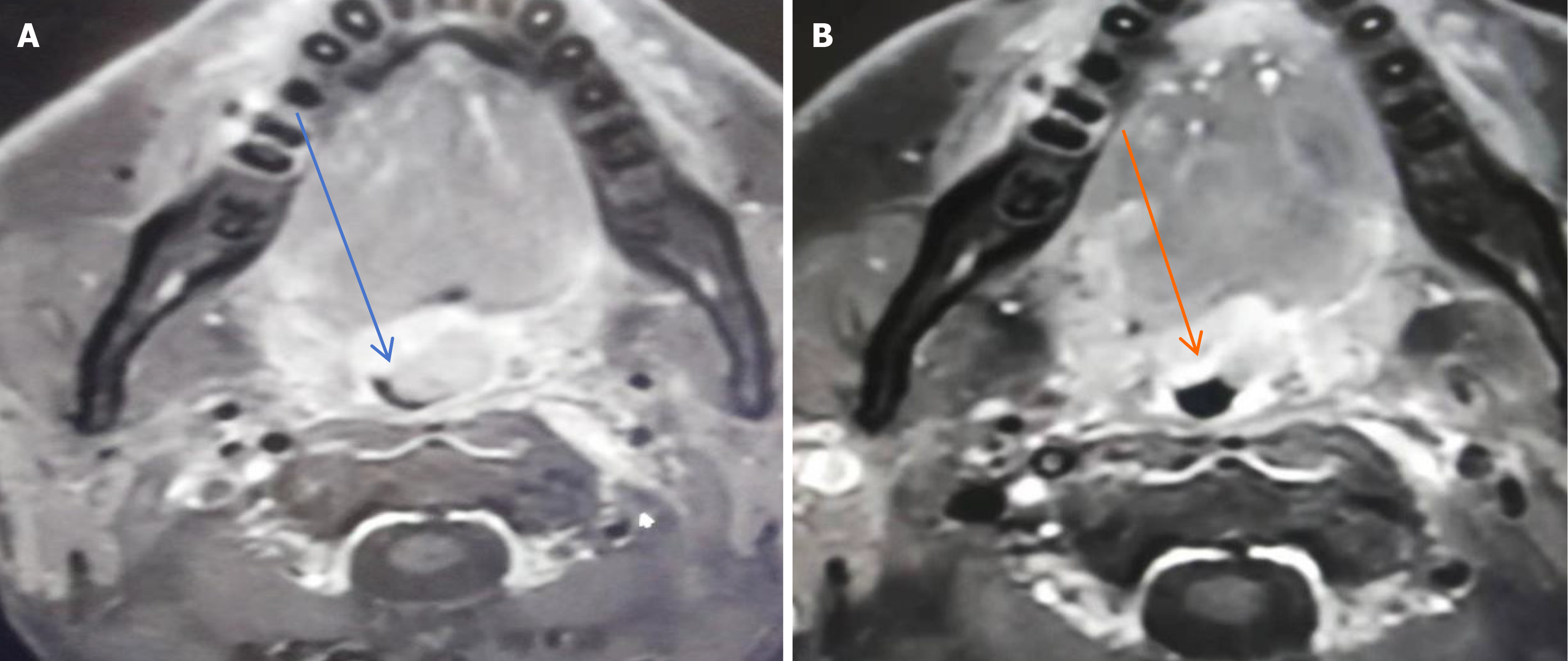
Figure 2 Magnetic resonance imaging images showing a uvula mass (orange arrow) and the uvula (blue arrow).
A: February 2023; B: April 2023.
- Citation: Yang J, Peng H, Tu SK, Li M, Song K. Extramedullary plasmacytoma with the uvula as first affected site: A case report. World J Clin Oncol 2025; 16(1): 96131
- URL: https://www.wjgnet.com/2218-4333/full/v16/i1/96131.htm
- DOI: https://dx.doi.org/10.5306/wjco.v16.i1.96131


