©The Author(s) 2024.
World J Clin Oncol. Sep 24, 2024; 15(9): 1157-1167
Published online Sep 24, 2024. doi: 10.5306/wjco.v15.i9.1157
Published online Sep 24, 2024. doi: 10.5306/wjco.v15.i9.1157
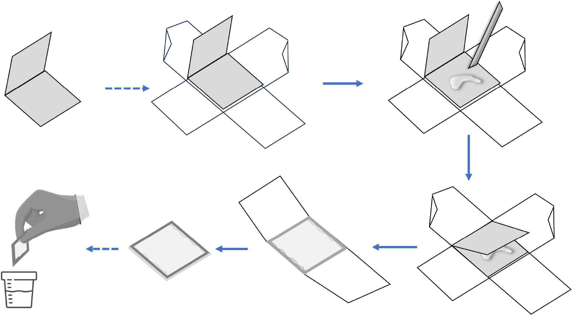
Figure 1 The modified protocol improved orientation gives pathologists a better representation representation of mucosal architecture and crypt bases.
In practice, the specimen envelope can be used in the procedure room during colonoscopy. Firstly, the envelope can be open on the counter top. After performing a polypectomy, the assistant transfers the specimen onto the envelope’s bottom flap, with two pieces of standard adhesive tape attached perpendicularly for easy retrieval. Use a toothpick to gently flatten the specimens, of which the mucosal surface would face up or down. The top flap was then gently folded over the polyp and taped shut. The tape completely encircled the envelope so that tape was affixed to tape. The perpendicular application prevented the specimen from falling out through the sides of the envelope. The corners of the envelope were left untaped, which allowed formalin to enter both the envelope and the specimen. Finally, this complex was placed in a jar of formalin.
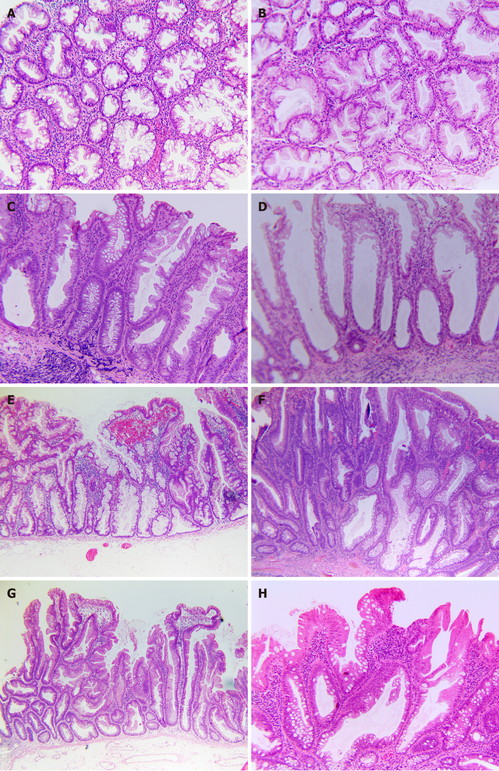
Figure 2 Chromatogram by general biopsy-specimen processing techniques.
A and B: Poorly orientated, horizontal sections (H&E, 100 ×); A: Case No. 1 with serrated pattern might require more information on polyp size and location to guide differential diagnosis between hyperplastic polyp (HP) and sessile serrated lesion (SSL); B: Case No. 2, which revealed hypercellularity and structure asymmetry, highly suggested a SSL with dysplasia (SSLD). Therefore, MLH1 staining was required for confirmative diagnosis; C and D: Crypt-base or crypt dilation might be hard to define in equivocal cases (H&E, 100 ×); C: Case No. 3 is rugged to evaluate for crypt dilation; however, the serrated feature could support diagnosis as a SSL; D: Case No. 4, which only showed the crypt base dilated without other features, could lead to being misdiagnosed or debated; E and F: Well-performed endoscopic submucosal dissection specimen with the reserved orientation of SSL and SSLD; E: Case No. 5, SSL, demonstrated the lateral growth along muscularis mucosae, creating an L or an inverted T, the most reliable feature (H&E, 40 ×); F: Case No. 6, SSLD, by World Health Organization 2019, reclassified as a mixture of serrated and intestinal dysplasia by Cenaj et al[23] and reclassified as dysplasia not specified by Liu et al[24], and reclassified as SSLDnos by American Journal of Clinical Pathology 2021 (100 ×); G and H: Several uncommon cases of serrated lesions; G: Case No. 7, which was flat overall, should be categorized as traditional serrated adenoma, transforming from an SSL rather than SSLD (H&E, 40 ×); H: Case No. 8, SSL, which had an eosinophilic surface change, should not be interpreted as cytological dysplasia (H&E, 100 ×).
- Citation: Tran TH, Nguyen VH, Vo DT. How to "pick up" colorectal serrated lesions and polyps in daily histopathology practice: From terminologies to diagnostic pitfalls. World J Clin Oncol 2024; 15(9): 1157-1167
- URL: https://www.wjgnet.com/2218-4333/full/v15/i9/1157.htm
- DOI: https://dx.doi.org/10.5306/wjco.v15.i9.1157


