©The Author(s) 2024.
World J Clin Oncol. Jul 24, 2024; 15(7): 953-960
Published online Jul 24, 2024. doi: 10.5306/wjco.v15.i7.953
Published online Jul 24, 2024. doi: 10.5306/wjco.v15.i7.953
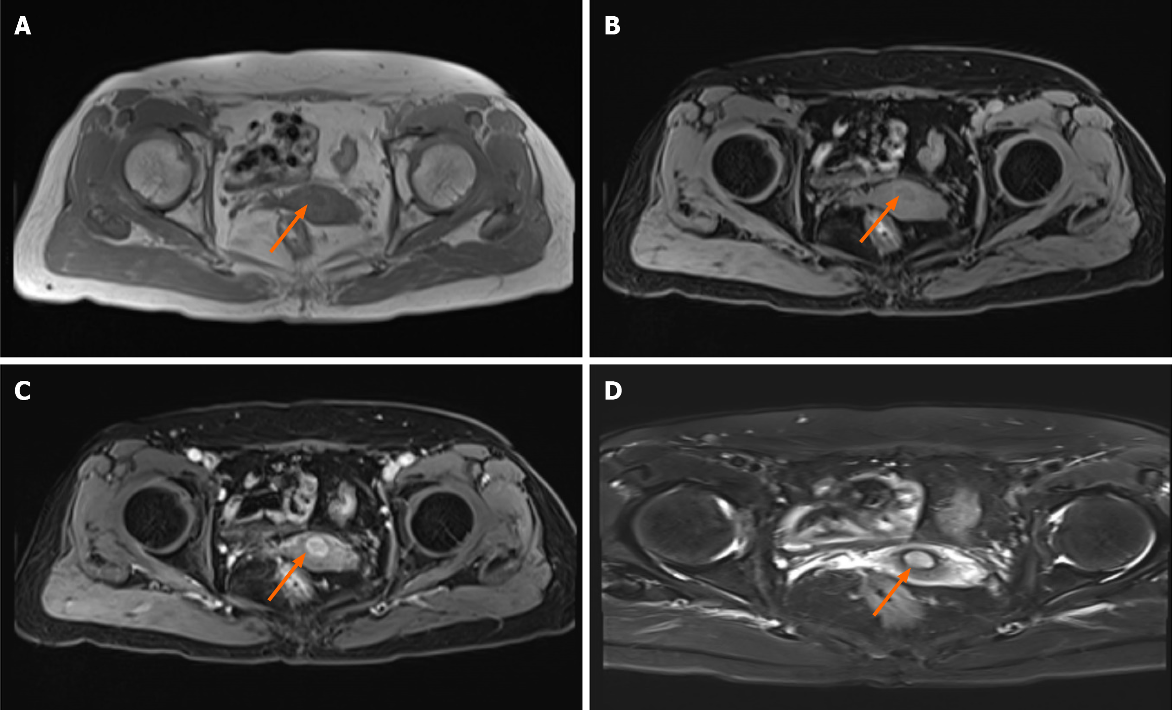
Figure 1 Pelvic magnetic resonance imaging.
A: T1 weighted image (arrow); B: Fat-suppression T1-weighted image (arrow); C: T1WI lipocompression enhanced scan (arrow); D: T2WI lipocompression enhanced scan (arrow).
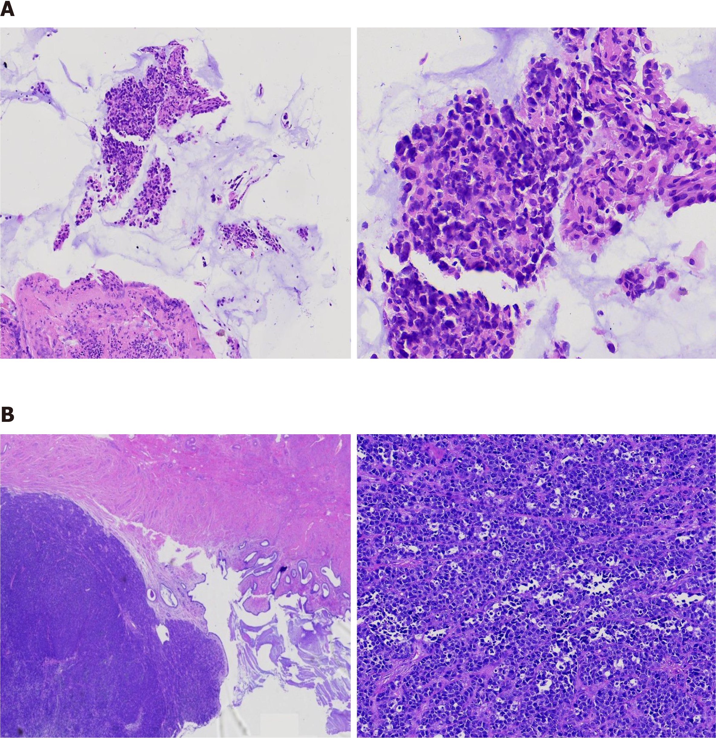
Figure 2 Histological examination showed small round cell malignancy with a high mitotic activity.
A: Colposcopy and cervical biopsy; B: Resected biopsy specimen.
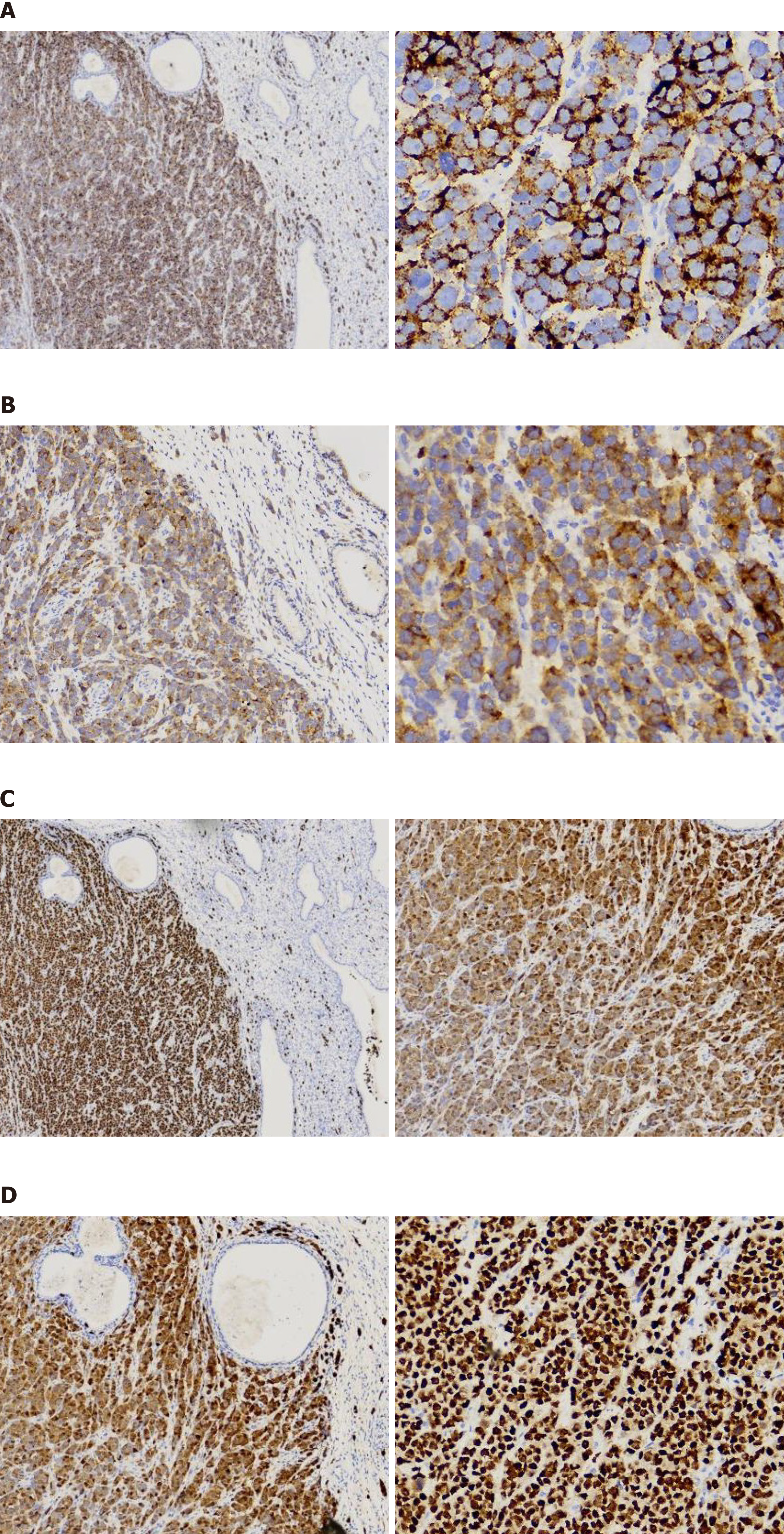
Figure 3 Immunohistochemical staining of resected biopsy specimen.
A: HMB45; B: MelanA; C: S-100; D: SOX10. The tumors were positive for HMB45, MelanA, S-100 and SOX10. Scale bar, left, 100 μm; right, 400 μm.
- Citation: Duan JL, Yang J, Zhang YL, Huang WT. Amelanotic primary cervical malignant melanoma: A case report and review of literature. World J Clin Oncol 2024; 15(7): 953-960
- URL: https://www.wjgnet.com/2218-4333/full/v15/i7/953.htm
- DOI: https://dx.doi.org/10.5306/wjco.v15.i7.953


