©The Author(s) 2024.
World J Clin Oncol. May 24, 2024; 15(5): 580-586
Published online May 24, 2024. doi: 10.5306/wjco.v15.i5.580
Published online May 24, 2024. doi: 10.5306/wjco.v15.i5.580
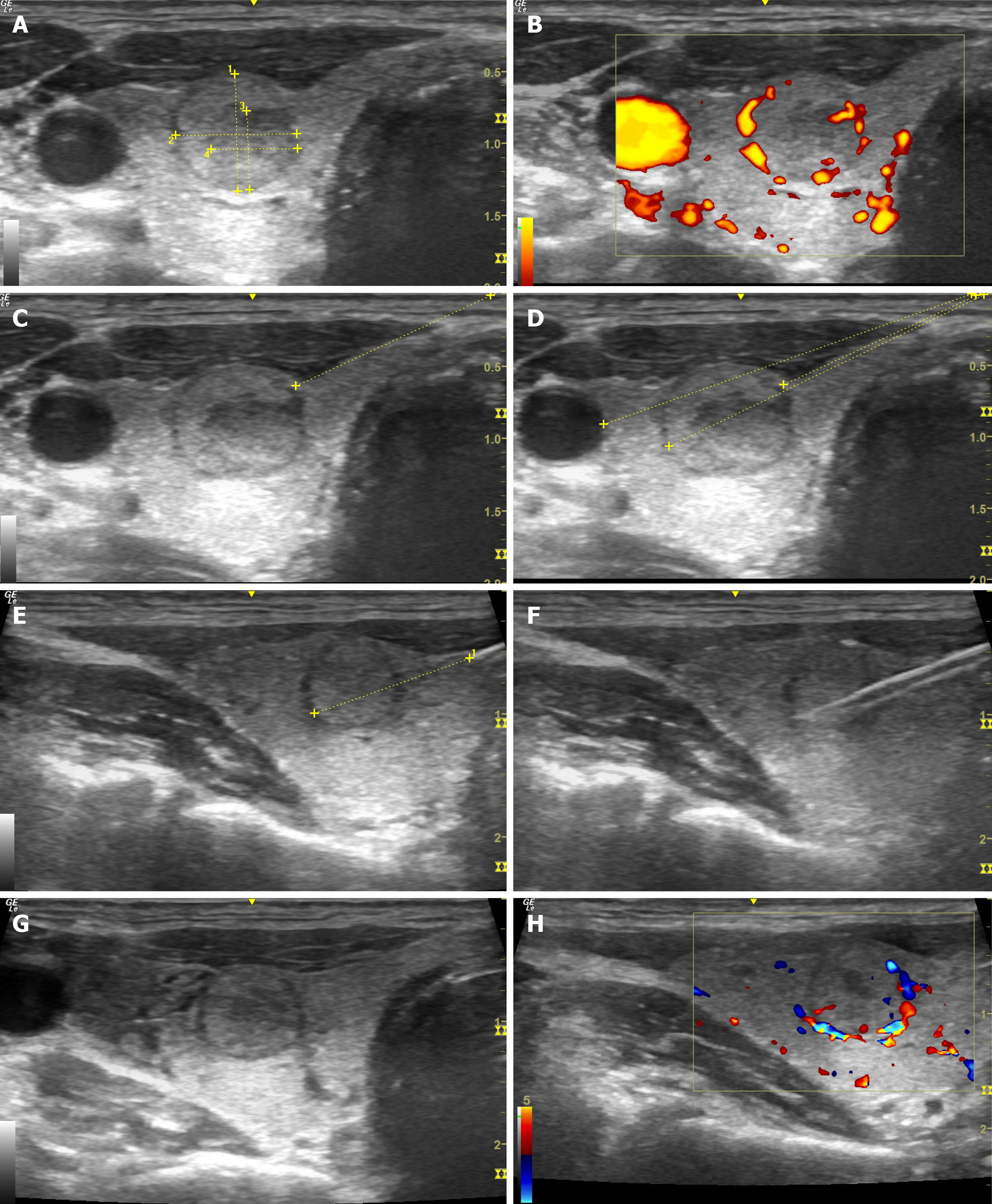
Figure 1 Ultrasonography-guided core-needle biopsy of the thyroid gland.
A: Assessment of the size of the mass; B: Assessment in the Doppler mode; C: Marking the optimal trajectory for core-needle biopsy (CNB); D: Calculation of the distance till the mass and major vessels; E: Control of the needle along its length; F: Control during the biopsy; G: Assessment of the mass after CNB; H: Assessment of the node in Doppler mode after CNB.
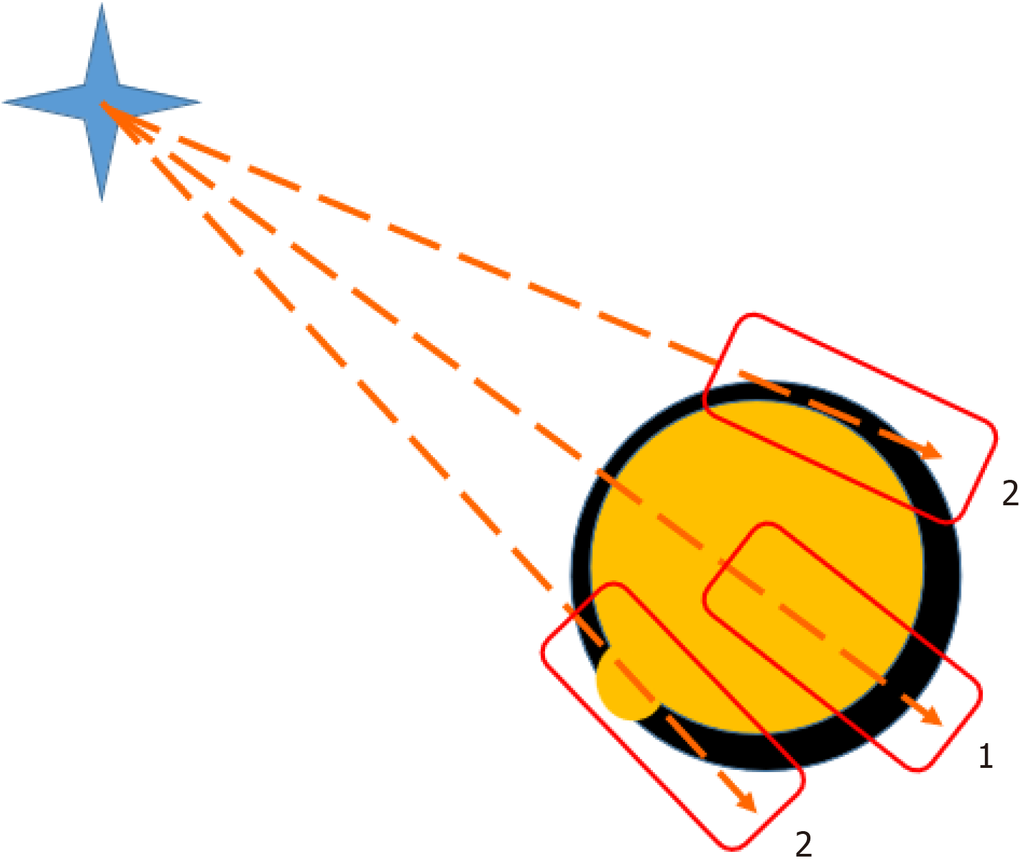
Figure 2
Core-needle biopsy of a thyroid node may be marginal 2 or through the node 1.
- Citation: Dolidze DD, Covantsev S, Chechenin GM, Pichugina NV, Bedina AV, Bumbu A. Core needle biopsy for thyroid nodules assessment-a new horizon? World J Clin Oncol 2024; 15(5): 580-586
- URL: https://www.wjgnet.com/2218-4333/full/v15/i5/580.htm
- DOI: https://dx.doi.org/10.5306/wjco.v15.i5.580


