©The Author(s) 2024.
World J Clin Oncol. Nov 24, 2024; 15(11): 1444-1453
Published online Nov 24, 2024. doi: 10.5306/wjco.v15.i11.1444
Published online Nov 24, 2024. doi: 10.5306/wjco.v15.i11.1444
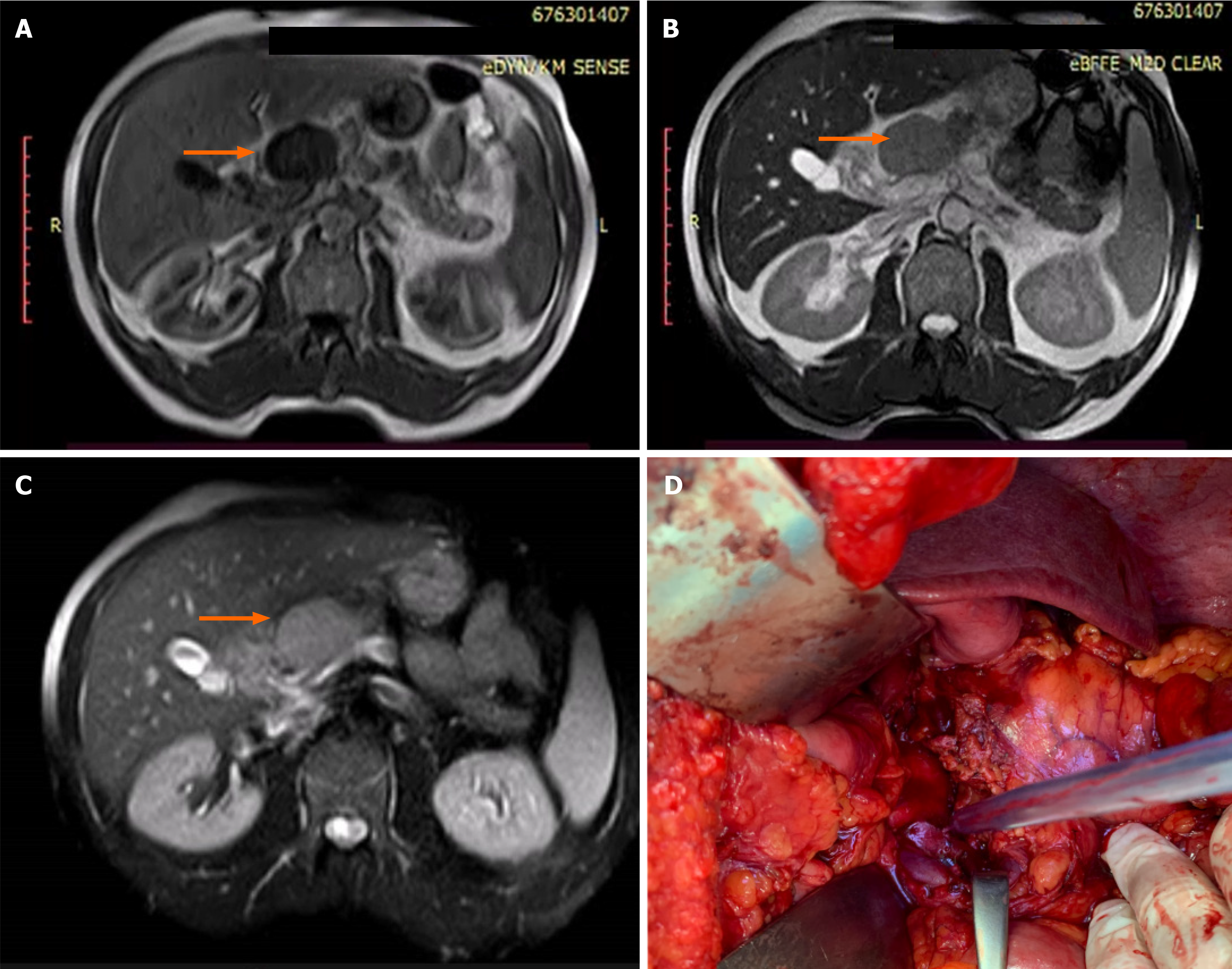
Figure 1 Magnetic resonance imaging and intraoperative finding of primary pancreatic lymphoma.
A: T1-weighted magnetic resonance imaging (MRI) revealed a well-circumscribed, oval, hypointense lesion (58 mm in diameter) located in the neck and body region; B: Lesion was hyperintense on the T2-weighted MRI; C: Slight post-contrast MRI enhancement; D: Intraoperative finding after resection of the pancreas.
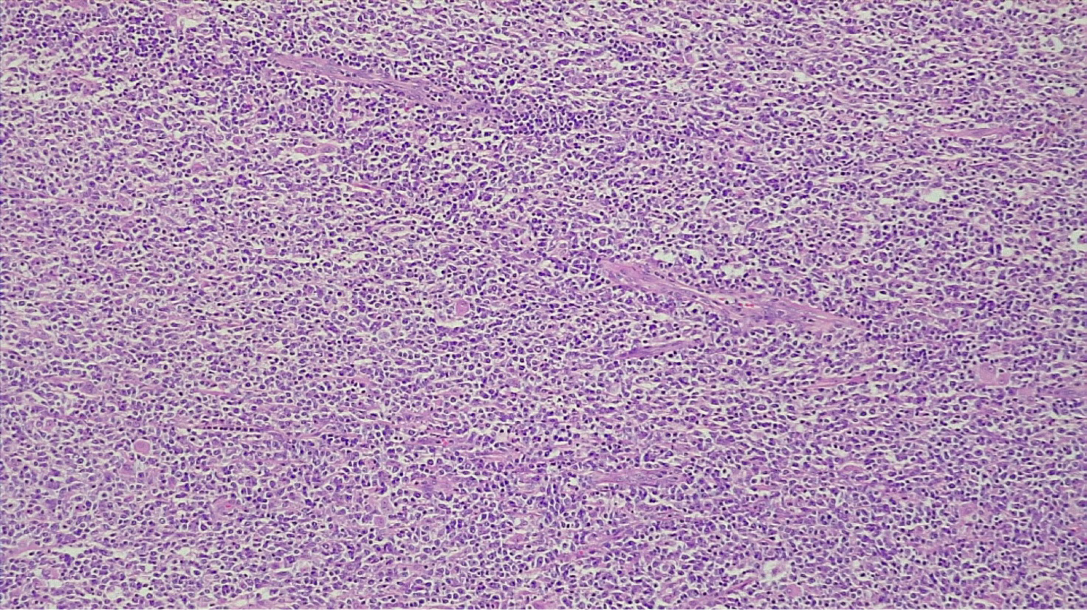
Figure 2 Hematoxylin and eosin staining revealed diffuse large B-cell lymphoma.
× 10 magnification.
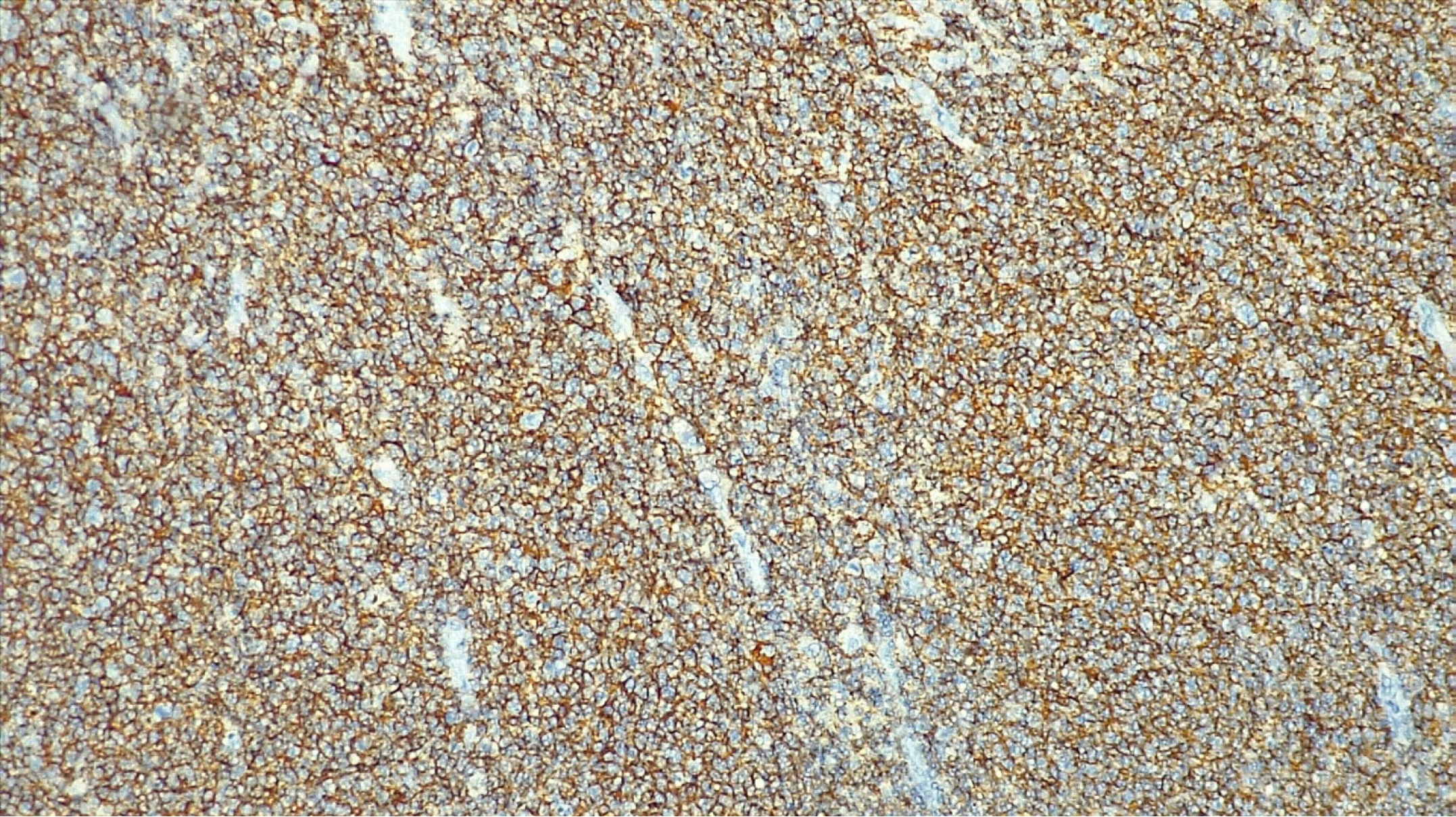
Figure 3 CD20 positivity.
× 10 magnification.
- Citation: Stojanovic MM, Brzacki V, Marjanovic G, Nestorovic M, Zivadinovic J, Krstic M, Gmijovic M, Golubovic I, Jovanovic S, Stojanovic MP, Terzic K. Primary pancreatic lymphoma: A case report and review of literature. World J Clin Oncol 2024; 15(11): 1444-1453
- URL: https://www.wjgnet.com/2218-4333/full/v15/i11/1444.htm
- DOI: https://dx.doi.org/10.5306/wjco.v15.i11.1444


