Copyright
©The Author(s) 2023.
World J Clin Oncol. Nov 24, 2023; 14(11): 471-478
Published online Nov 24, 2023. doi: 10.5306/wjco.v14.i11.471
Published online Nov 24, 2023. doi: 10.5306/wjco.v14.i11.471
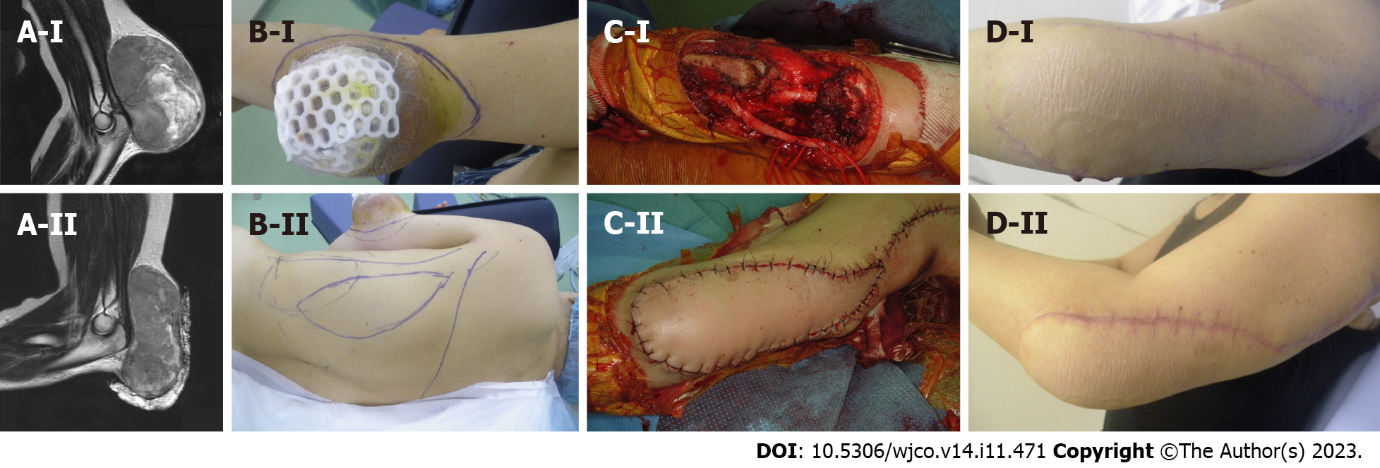
Figure 1 Synovial sarcoma at the distal upper arm (elbow) reconstructed by a pedicled latissimus dorsi (pedicled flap, CpDp).
A: A 47-year-old female with a synovial sarcoma at the elbow (distal upper arm). Magnetic resonance imaging showed a tumor with heterogenous low-to-high signal intensity on the T2-weighted image. Before (A-I) and after (A-II) chemotherapy of doxorubicin and ifosfamide, the tumor size was reduced. B-D: A wide surgical resection was performed with a pedicled latissimus dorsi. The CD-grade was CpDp (pedicled flap, CpDp).
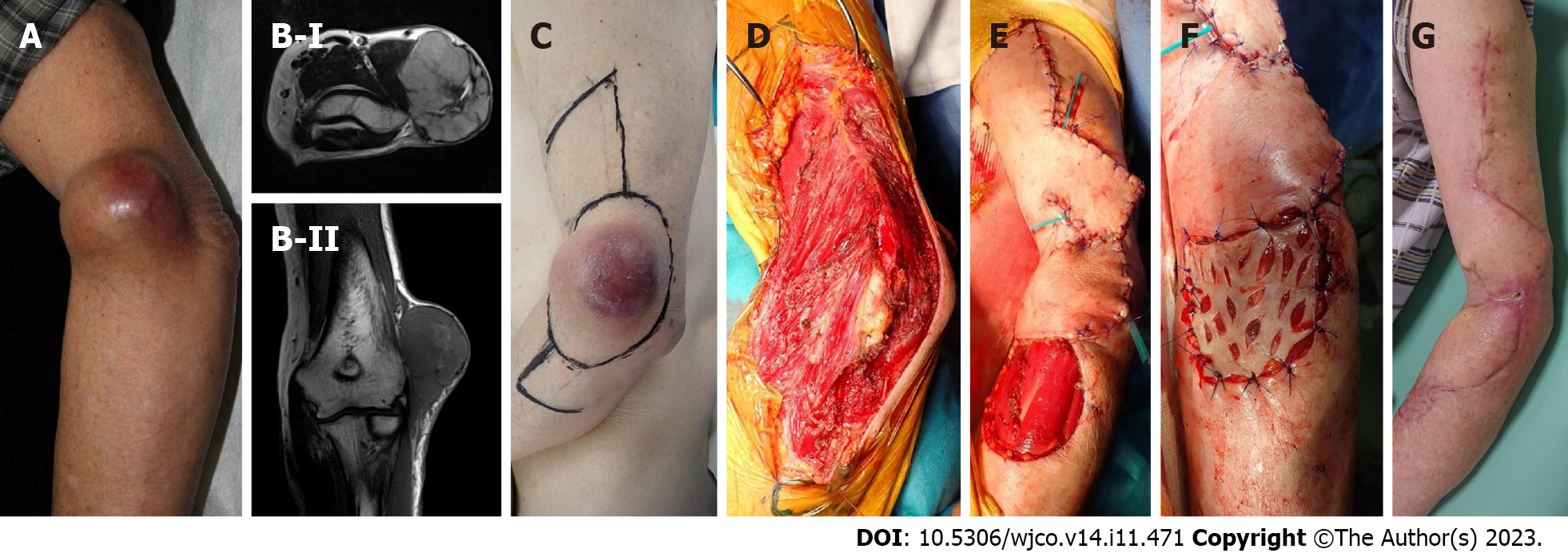
Figure 2 Pleomorphic rhabdomyosarcoma at the distal upper arm (elbow) reconstructed by transpositional flap (transpositional flap, CdD0).
A: An 85-year-old male with a pleomorphic rhabdomyosarcoma at the elbow (distal upper arm); B: Magnetic resonance imaging showed a tumor with homogenous high-signal intensity on T2-weighted images (B-I) and low-signal intensity on T1-weighted images (B-II). A wide surgical resection was performed. The transpositional flap was obtained from the upper arm and forearm; C-G: Skin grafting was performed at the forearm. The CD-grade was CdD0 (transpositional flap, CdD0).
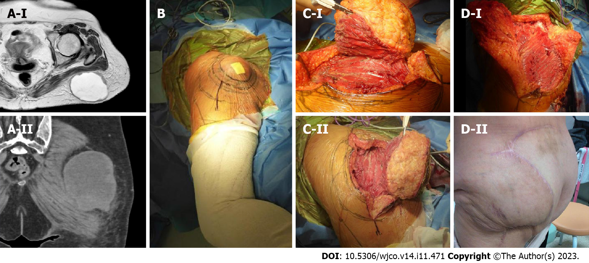
Figure 3 Undifferentiated pleomorphic sarcoma at the buttock reconstructed by a transpositional flap (transpositional flap, C0D0).
A: A 65-year-old female with an undifferentiated pleomorphic sarcoma at the buttock. Magnetic resonance imaging revealed a subcutaneous tumor. The tumor had a cystic appearance and contained liquid with slightly high signal intensity on the T2-weighted image. The periphery of the cystic wall was thick with a solid neoplastic lesion and intermediate signal intensity on T2-weighted images (A-I). Computed tomography showed that the lesion is located at the buttock (A-II); B: A resection of the tumor was designed; C and D: The tumor was resected and the defect was reconstructed with a transpositional flap donated from the lateral abdomen. The CD-grade was C0D0 (transpositional flap, C0D0).
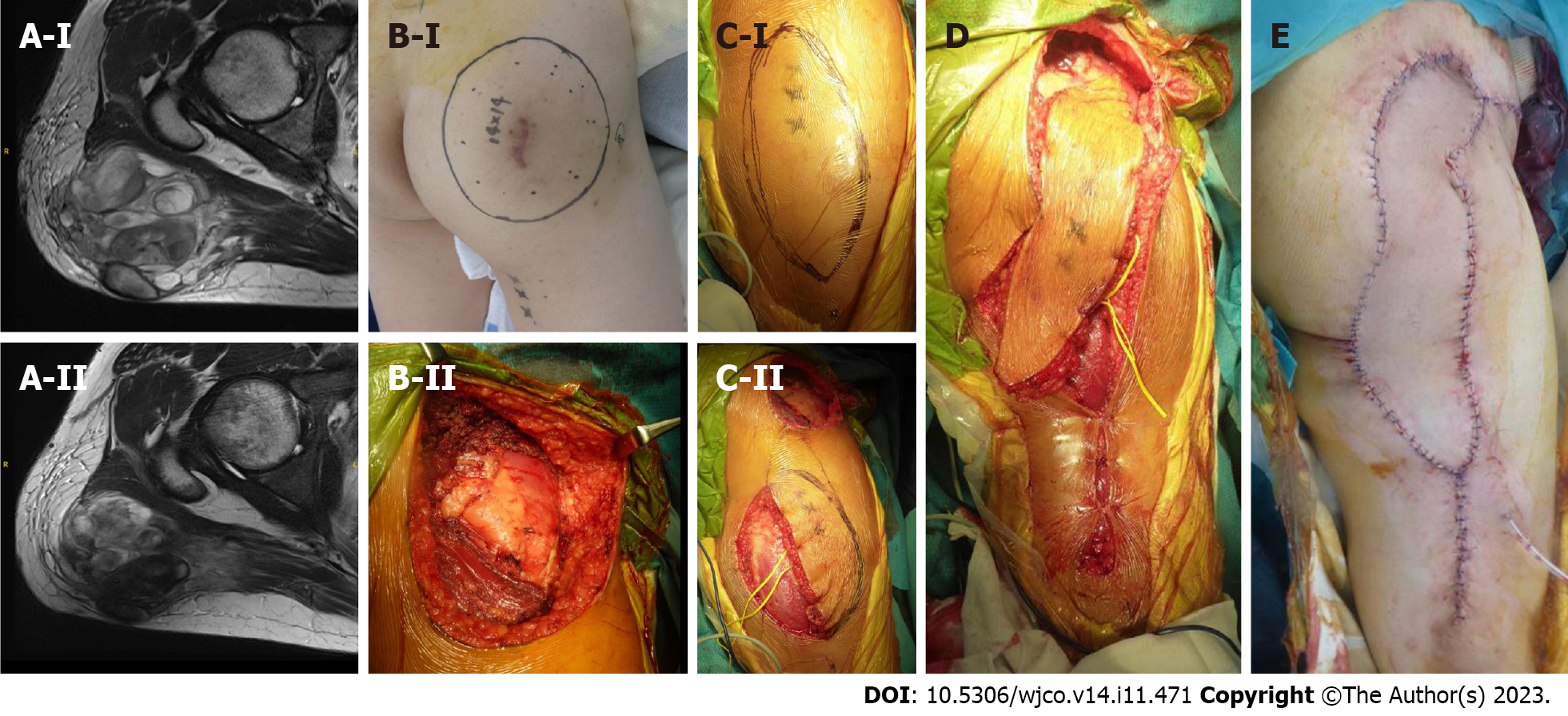
Figure 4 Myxofibrosarcoma at the buttock reconstructed by a propeller flap (pedicled flap, CdDd).
A: A 46-year-old male with a myxofibrosarcoma at the buttock. Magnetic resonance imaging revealed that the tumor showed heterogenous low-to-high signal intensity on the T2-weighted image. Before (A-I) and after (A-II) chemotherapy of doxorubicin and ifosfamide, the tumor size was reduced; B: The resection of the tumor was designed (B-I) and performed (B-II); C and E: A propeller flap from the thigh was designed (C-I) and the pedicle was preserved (C-II) and performed. The CD-grade was CdDd (pedicled flap, CdDd).
- Citation: Sakamoto A, Noguchi T, Matsuda S. System describing surgical field extension associated with flap reconstruction after resection of a superficial malignant soft tissue tumor. World J Clin Oncol 2023; 14(11): 471-478
- URL: https://www.wjgnet.com/2218-4333/full/v14/i11/471.htm
- DOI: https://dx.doi.org/10.5306/wjco.v14.i11.471


