©The Author(s) 2022.
World J Clin Oncol. Apr 24, 2022; 13(4): 287-302
Published online Apr 24, 2022. doi: 10.5306/wjco.v13.i4.287
Published online Apr 24, 2022. doi: 10.5306/wjco.v13.i4.287
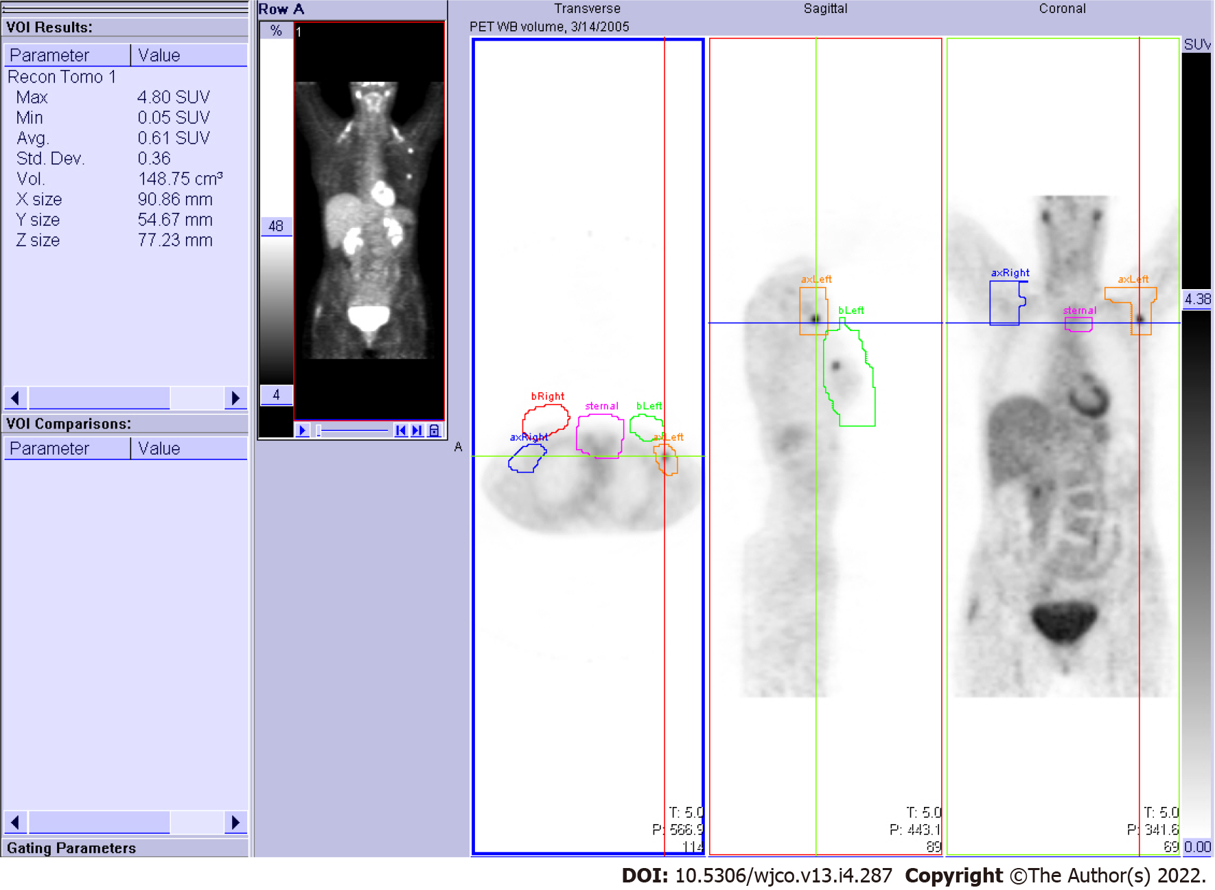
Figure 1 Anatomical regions of interest.
Free-hand three-dimensional non-overlapping anatomical regions of interest (AROI) volumes drawn on the PET scan workstation for the right breast (red), right axilla-supraclavicular nodal area (blue), left breast (green), left axilla-supraclavicular (orange), and sternal-mediastinal area (purple). Viewing planes are indicated by crosshairs.
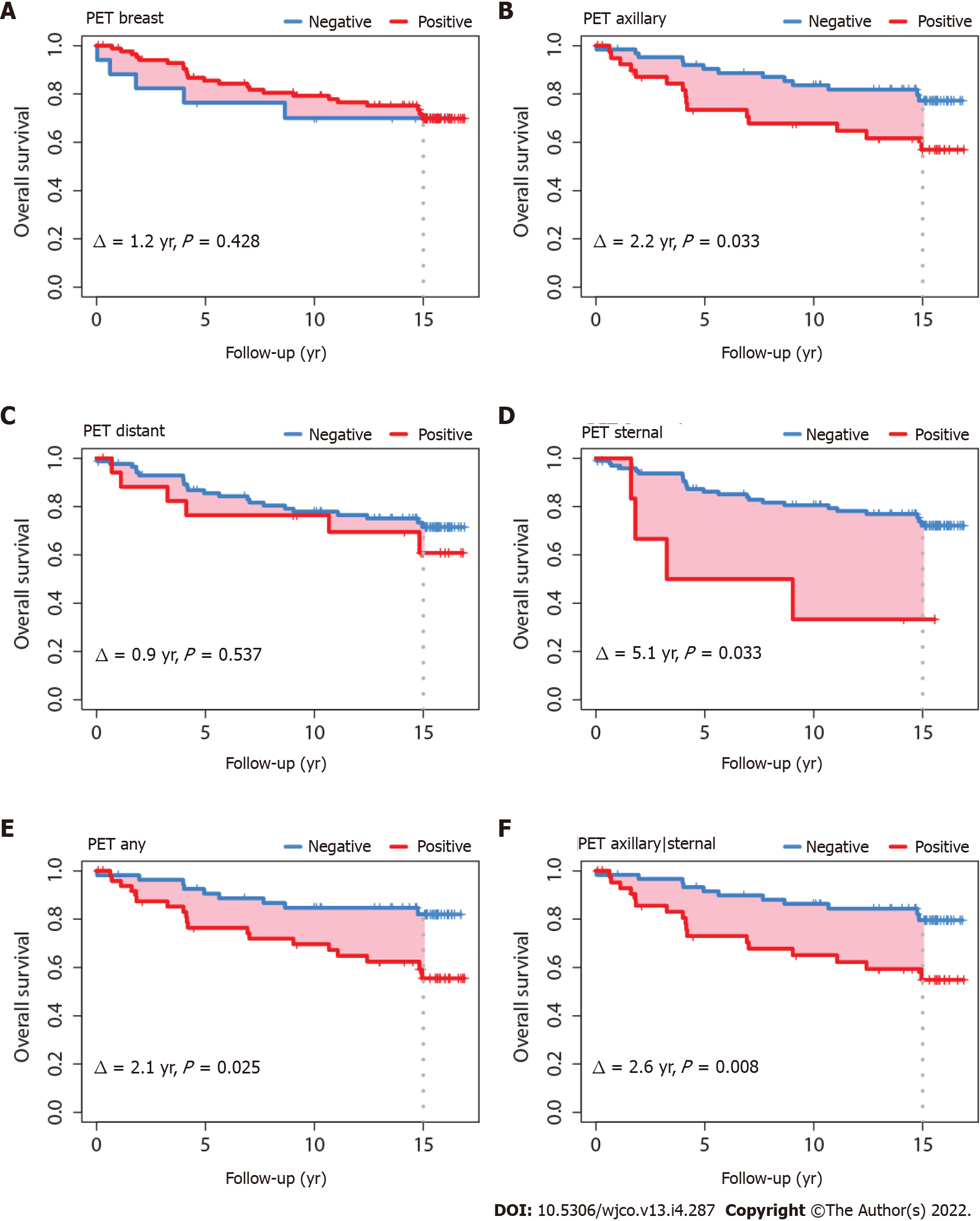
Figure 2 Overall survival according to fluorine-18-fluorodeoxyglucose positron-emission tomography status in anatomical regions of interest.
A: Breast; B: Axillary; C: Distant; D: Sternal; E: Any axillary, sternal or distant; F: Any axillary or sternal.
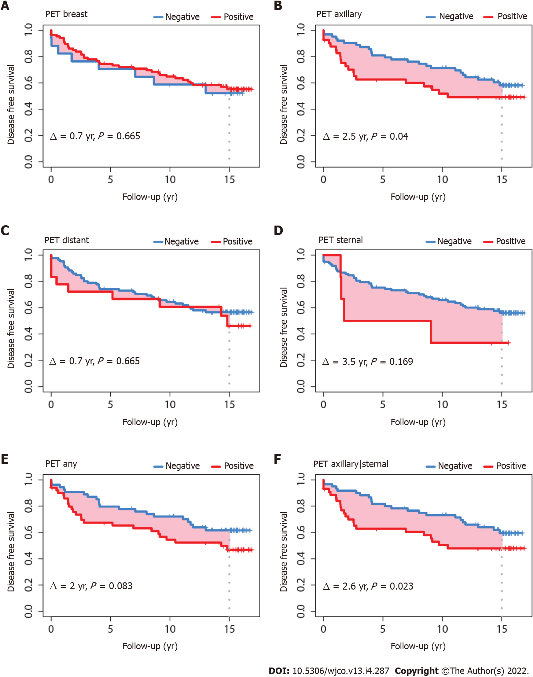
Figure 3 Disease-free survival according to fluorine-18-fluorodeoxyglucose positron-emission tomography status in anatomical regions of interest.
A: Breast; B: Axillary; C: Distant; D: Sternal; E: Any axillary, sternal or distant; F: Any axillary or sternal.
- Citation: Perrin J, Farid K, Van Parijs H, Gorobets O, Vinh-Hung V, Nguyen NP, Djassemi N, De Ridder M, Everaert H. Is there utility for fluorine-18-fluorodeoxyglucose positron-emission tomography scan before surgery in breast cancer? A 15-year overall survival analysis. World J Clin Oncol 2022; 13(4): 287-302
- URL: https://www.wjgnet.com/2218-4333/full/v13/i4/287.htm
- DOI: https://dx.doi.org/10.5306/wjco.v13.i4.287


