©The Author(s) 2021.
World J Clin Oncol. Dec 24, 2021; 12(12): 1227-1243
Published online Dec 24, 2021. doi: 10.5306/wjco.v12.i12.1227
Published online Dec 24, 2021. doi: 10.5306/wjco.v12.i12.1227
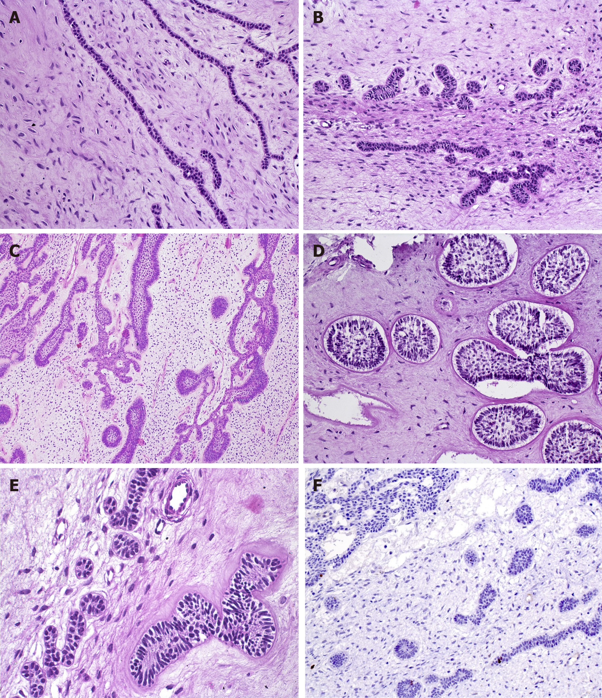
Figure 1 Diverse aspects of the odontogenic epithelium in ameloblastic fibromas within the cell-rich myxoid stroma.
A: Epithelial strands, comprising a double layer of cuboidal cells (HE, 20×); B: Epithelial proliferation with primitive appearance that resembles tooth bud-like structures (HE, 20×); C: Epithelial component with a follicular pattern comprising columnar cells at the periphery of the nests with central stellate reticulum-like cells (HE, 10×); D: Clefts of mesenchymal tissue surrounding follicular epithelial proliferations (HE, 20×); E: Mild hyalinization surrounding the basal layer of the epithelial nest (left). Smaller epithelial rosette-like islands resemble remnants of dental lamina (right) (HE, 40×); F: A very low rate of proliferation in both mesenchymal and epithelial components, showing the benign behavior of ameloblastic fibromas (IHC for Ki-67, 20×).
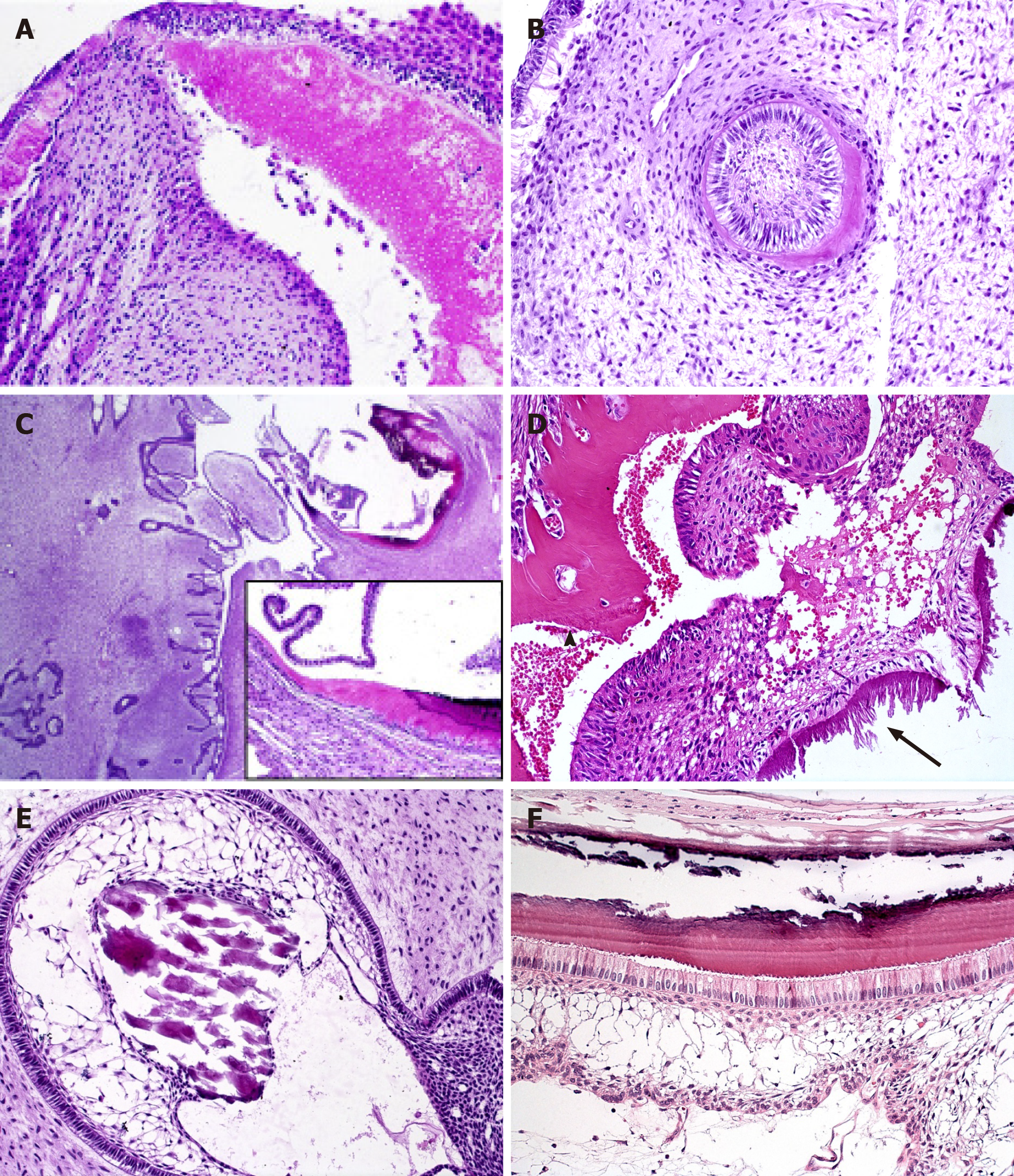
Figure 2 Mineralized tissue formation in ameloblastic fibrodentinoma and ameloblastic fibro-odontoma.
A, B: Dentinoid induction by epithelial cells in ameloblastic fibrodentinoma; note the presence of tubules in (A) (HE, 20×); C: Prominent proliferation of soft tissue similar to ameloblastic fibromas and focal areas of dentinoid and enamel matrix production in close relationship with the epithelial component in ameloblastic fibro-odontoma (HE, 2.5×; inset 20×); D: Structures similar to tubules are observed in the dentinoid (arrowhead), which can be associated with odontogenic epithelium or ectomesenchymal tissue, while enamel matrix (arrow) associated with columnar odontogenic appears more basophilic, with different patterns of deposition that can resemble prisms or globules (HE, 20×); E: Calcificated material, compatible with enameloid, in direct relationship with epithelial cells of the stellate reticulum-like area (HE, 20×); F: Details of the columnar ameloblast-like cells with reverse polarization producing enamel matrix in which the “fish scale” pattern is visible. Flattened cells between the columnar cells and stellate reticulum-like area resemble the stratum intermedium of the tooth germ, which is believed to assist the ameloblast in producing enamel during odontogenesis (HE, 40×).
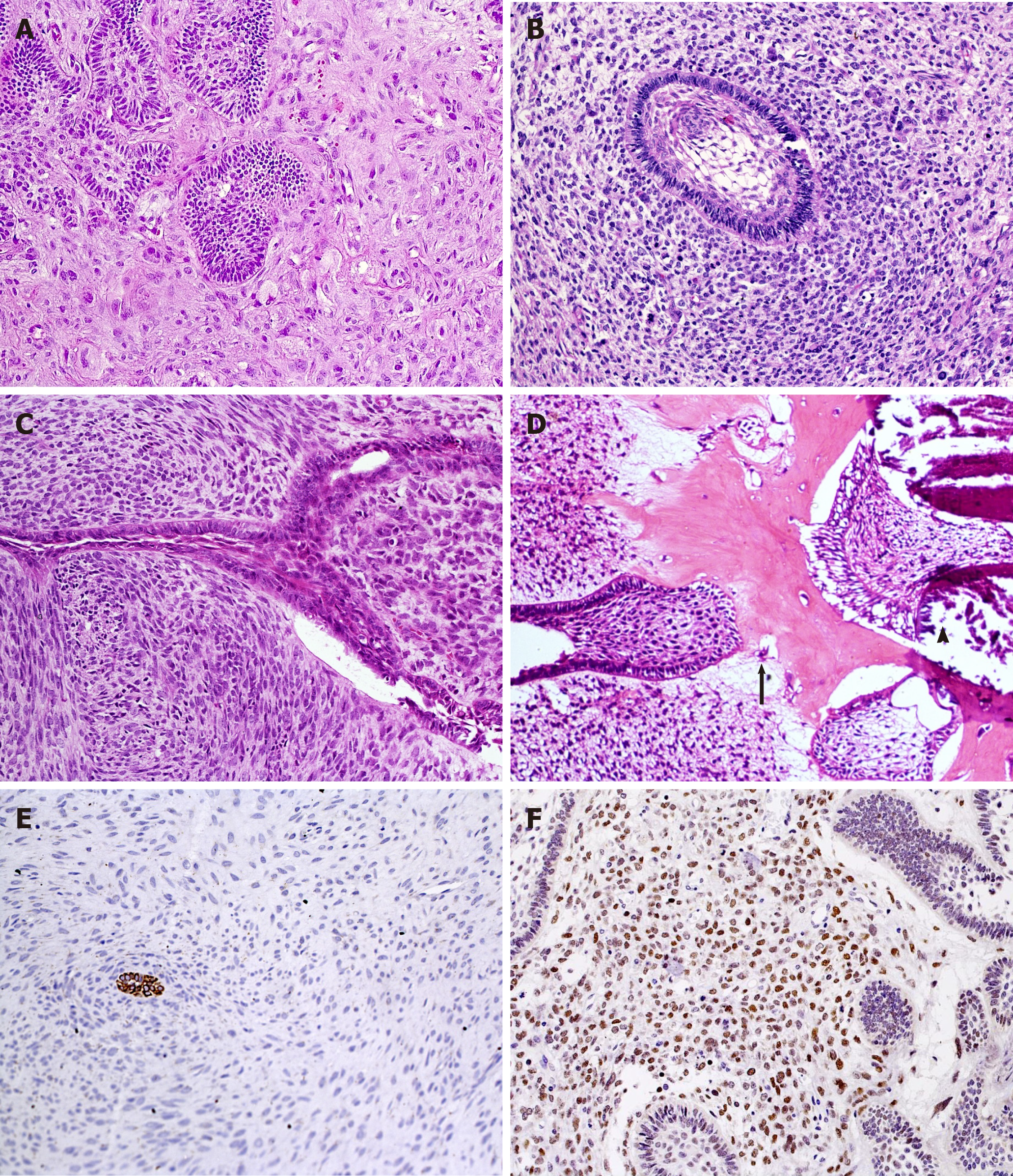
Figure 3 Histopathological aspects of ameloblastic fibromas and ameloblastic fibro-odontosarcoma.
Marked pleomorphism and atypia in mesenchymal cells (HE, 20×) (A-C). A: Several mitotic figures, nuclear hyperchromatism and multinucleated and aberrant cells are seen in a highly pleomorphic sarcomatous component of ameloblastic fibromas, while epithelial islands remain benign; B: A follicular benign epithelial island is surrounded by hypercellularized sarcomatous proliferation; C: Malignant mesenchymal tissue resembling a storiform pattern and haphazard disposition of sarcomatous cells around an epithelial branching cord; D: Production of enamel matrix (arrowhead) and dentinoid (arrow) as well as malignant mesenchymal tissue (left side) are components of this ameloblastic fibro-odontosarcoma (A-D: HE, 20×); E: Cytokeratins can help to localize odontogenic epithelial cells within dominant sarcomatous proliferation (IHC for AE1/AE3, 20×); F: Most mesenchymal malignant cells show nuclear positivity for p53 antigen (IHC for p53, 20×).
- Citation: Sánchez-Romero C, Paes de Almeida O, Bologna-Molina R. Mixed odontogenic tumors: A review of the clinicopathological and molecular features and changes in the WHO classification. World J Clin Oncol 2021; 12(12): 1227-1243
- URL: https://www.wjgnet.com/2218-4333/full/v12/i12/1227.htm
- DOI: https://dx.doi.org/10.5306/wjco.v12.i12.1227


