©The Author(s) 2021.
World J Clin Oncol. Nov 24, 2021; 12(11): 1083-1088
Published online Nov 24, 2021. doi: 10.5306/wjco.v12.i11.1083
Published online Nov 24, 2021. doi: 10.5306/wjco.v12.i11.1083
Figure 1 The necrotic ileum was resected, and the right femoral hernia was repaired using the McVay procedure.
A: The ileum was incarcerated into the right femoral hernia; B: The incarcerated ileum became necrotic; C: The resected ileum showed hemorrhage necrosis over a 10 cm length.
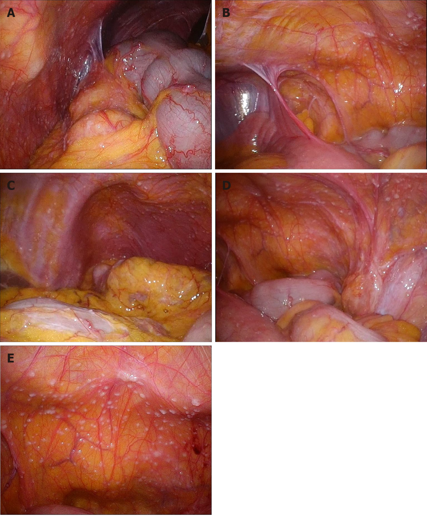
Figure 2 Multiple small white nodules mimicking peritoneal dissemination were observed throughout the entire abdominal cavity during the second operation.
A: Right upper abdominal cavity; B: Right lower abdominal cavity; C: Left upper abdominal cavity; D: Left lower abdominal cavity; E: Back side of the bladder.
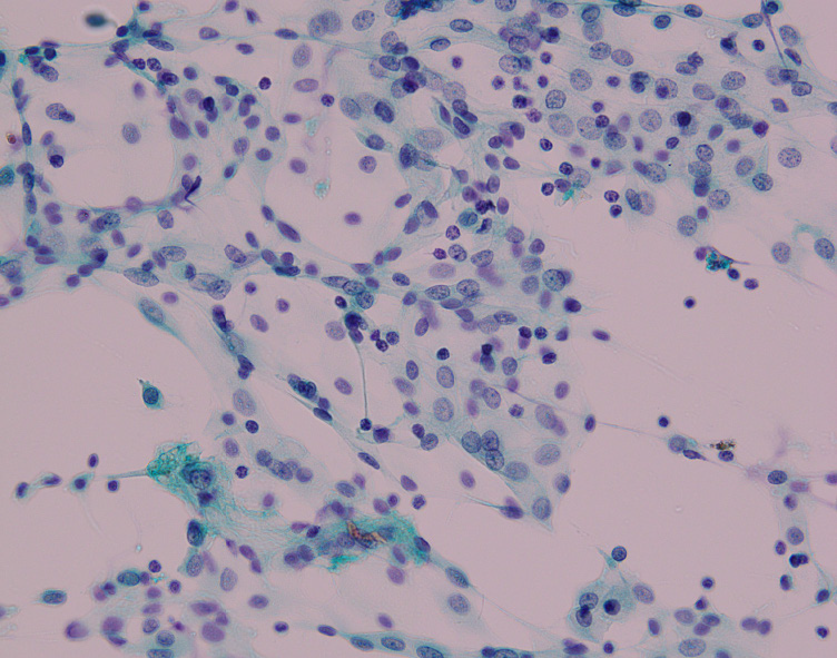
Figure 3 Papanicolaou stain of the peritoneal washing cytology of Douglas’ pouch showed only inflammatory cells (× 40).
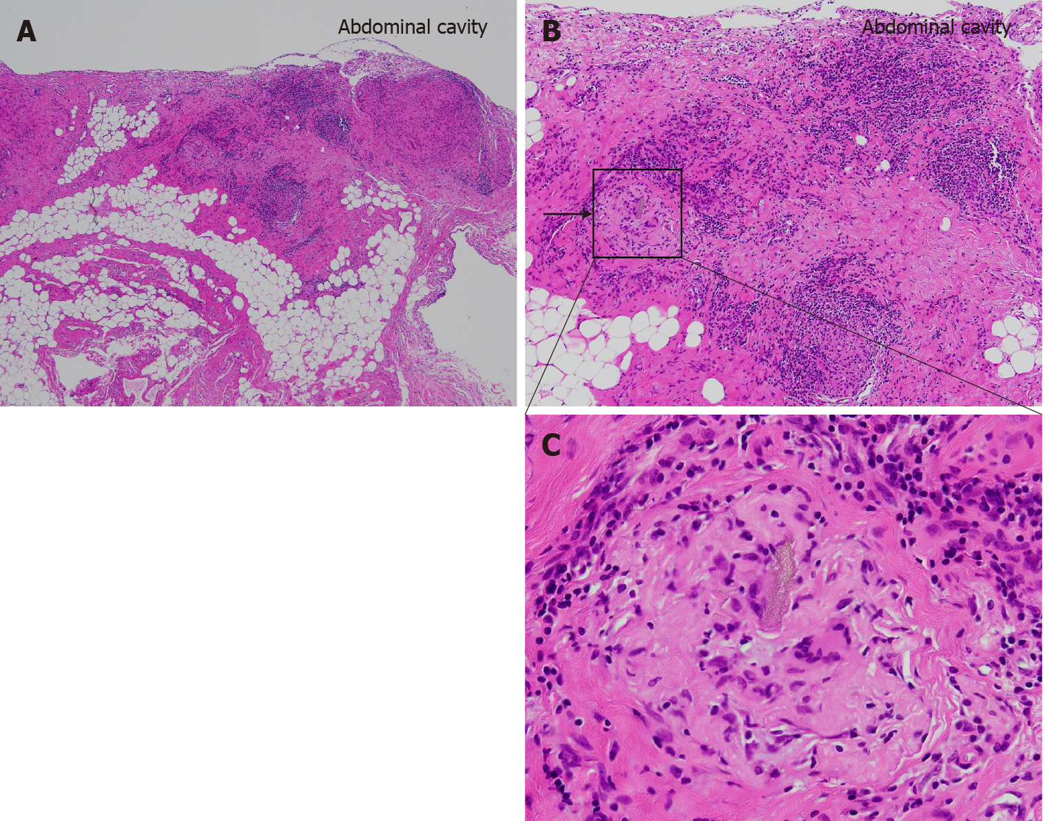
Figure 4 Pathological findings of the small white nodule.
Hematoxylin and eosin stain of the nodule. A multinucleated giant cell with a foreign body (arrow) surrounded by monocytes, lymphocytes, and granuloma formations. A: × 40; B: × 100; C: × 200.
- Citation: Ogino S, Matsumoto T, Kamada Y, Koizumi N, Fujiki H, Nakamura K, Yamano T, Sakakura C. Foreign body granulomas mimic peritoneal dissemination caused by incarcerated femoral hernia perforation: A case report. World J Clin Oncol 2021; 12(11): 1083-1088
- URL: https://www.wjgnet.com/2218-4333/full/v12/i11/1083.htm
- DOI: https://dx.doi.org/10.5306/wjco.v12.i11.1083


