Copyright
©The Author(s) 2020.
World J Clin Oncol. Apr 24, 2020; 11(4): 243-249
Published online Apr 24, 2020. doi: 10.5306/wjco.v11.i4.243
Published online Apr 24, 2020. doi: 10.5306/wjco.v11.i4.243
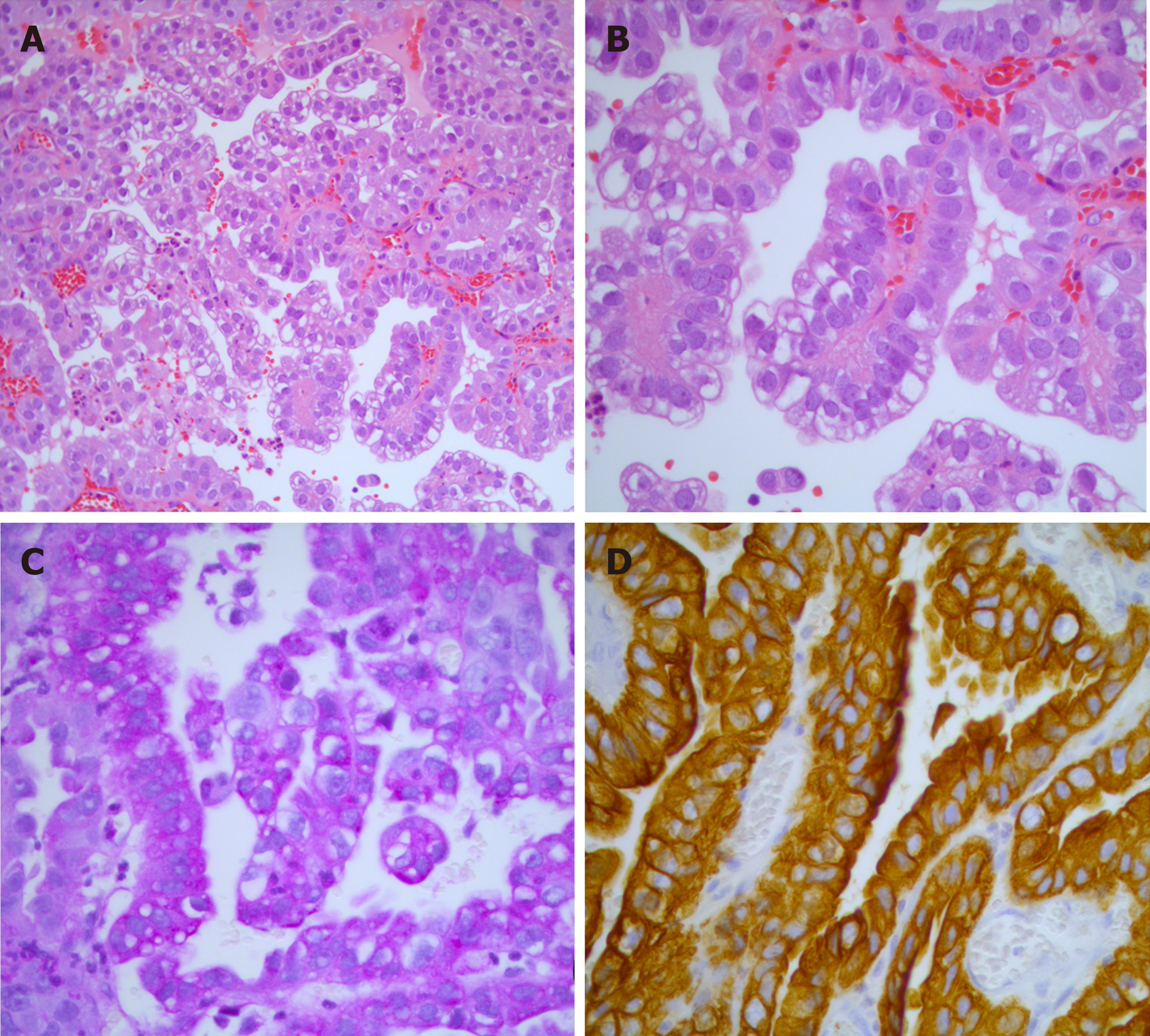
Figure 1 Histopathological examination.
A: Histological examination of clear cell adenoma of the urethra demonstrating the tubulopapillary growth pattern of the tumor (Hematoxylin-eosin staining, 200 ×); B: Higher magnification view of (A), with a better view of the clear cytoplasm and cytologic details (Hematoxylin-eosin staining, 400 ×); C: Tumor cells with intracytoplasmic periodic acid-schiff-positive material, consistent with glycogen (periodic acid-schiff stain, 400 ×); D: Tumor cells are diffusely positive for the epithelial marker cytokeratin 7 (Cytokeratin 7 Immunohistochemistry stain, 400 ×).
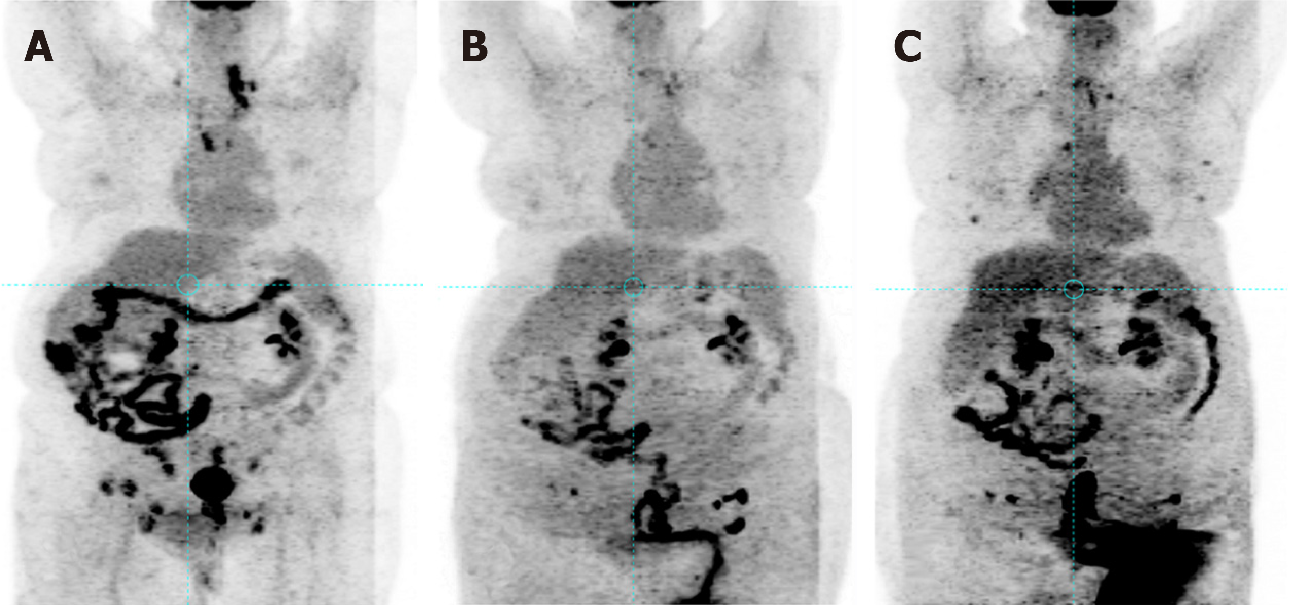
Figure 2 Positron emission tomography scan.
A: Prior to non-platinum-based chemotherapy consisting of paclitaxel/bevacizumab; B: Following 3 cycles of this combination chemotherapy; C: After 6 cycles of this combination chemotherapy.
- Citation: Shields LBE, Kalebasty AR. Personalized chemotherapy in clear cell adenocarcinoma of the urethra: A case report. World J Clin Oncol 2020; 11(4): 243-249
- URL: https://www.wjgnet.com/2218-4333/full/v11/i4/243.htm
- DOI: https://dx.doi.org/10.5306/wjco.v11.i4.243


