©The Author(s) 2020.
World J Clin Oncol. Dec 24, 2020; 11(12): 1070-1075
Published online Dec 24, 2020. doi: 10.5306/wjco.v11.i12.1070
Published online Dec 24, 2020. doi: 10.5306/wjco.v11.i12.1070
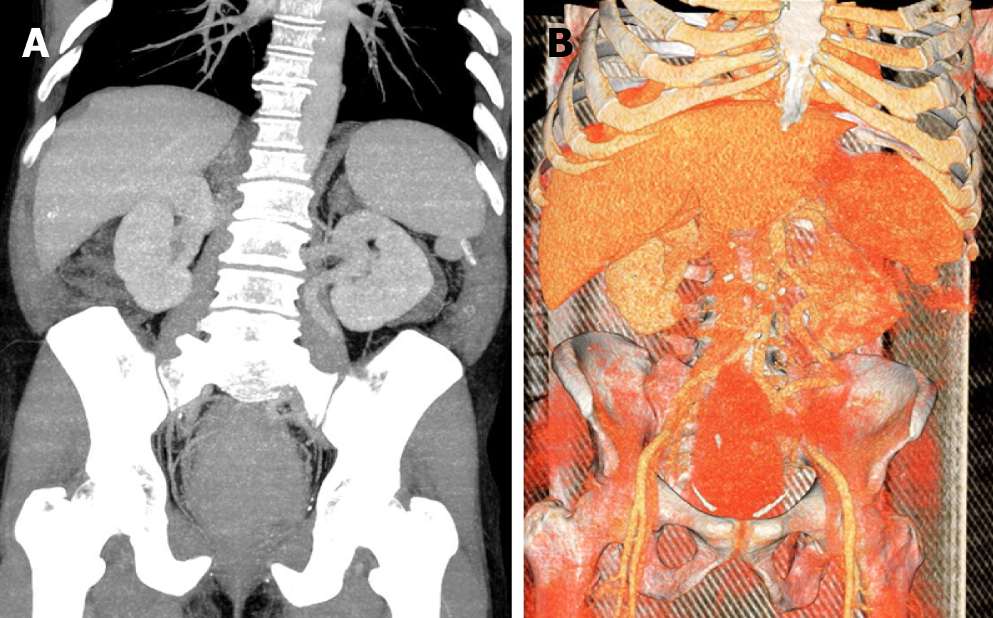
Figure 1 Computed tomography scans with vascular reconstruction.
A: A bulky rectal tumor with B: Marked hypervascularization.
Figure 2 Preoperative embolization.
A: Digital subtraction angiography (DSA) of the inferior mesenteric artery identifying the enlarged superior rectal artery with prominent branches supplying the rectal tumor; B: DSA of the inferior mesenteric artery after 300-500 micra tris-acryl gelatin microspheres distal embolization into the superior rectal artery and partial occlusion of its main branch with controlled detachable platinum coils; C: Final DSA control of the inferior mesenteric artery after injection of N-butyl-2 cyanoacrylate with ethiodol 1:4 at the bifurcation of the right and left branches of the superior rectal artery, showing a significant reduction in distal arterial supply.
Figure 3 Surgical specimen after abdominal perineal resection.
A: Total mesorectal excision; B: Extensive tumor necrosis can be observed after opening the surgical specimen.
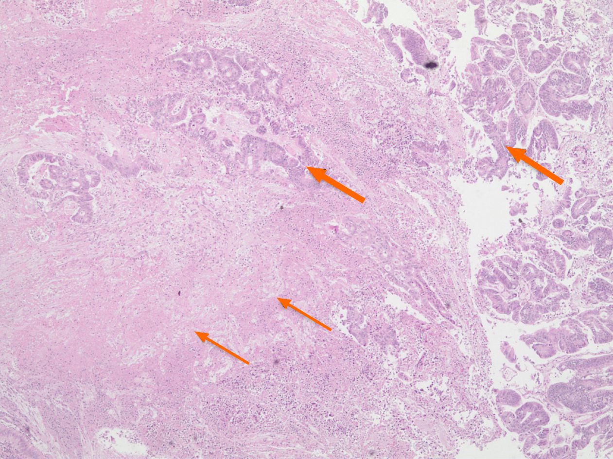
Figure 4 Rectal adenocarcinoma (thick arrows) showing extensive tumor necrosis (thin arrows).
Hematoxylin and Eosin stain, × 40 magnification.
- Citation: Feitosa MR, de Freitas LF, Filho AB, Nakiri GS, Abud DG, Landell LM, Brunaldi MO, da Rocha JJR, Feres O, Parra RS. Preoperative rectal tumor embolization as an adjunctive tool for bloodless abdominoperineal excision: A case report. World J Clin Oncol 2020; 11(12): 1070-1075
- URL: https://www.wjgnet.com/2218-4333/full/v11/i12/1070.htm
- DOI: https://dx.doi.org/10.5306/wjco.v11.i12.1070


