©The Author(s) 2020.
World J Gastrointest Pharmacol Ther. Aug 8, 2020; 11(3): 48-58
Published online Aug 8, 2020. doi: 10.4292/wjgpt.v11.i3.48
Published online Aug 8, 2020. doi: 10.4292/wjgpt.v11.i3.48
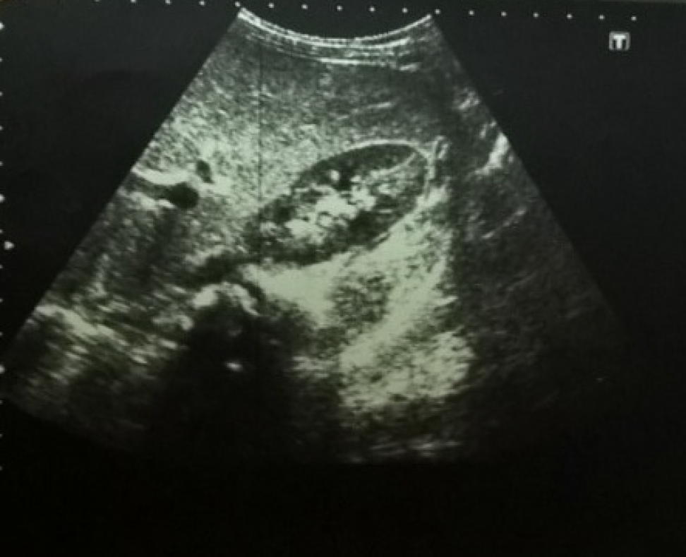
Figure 1 Abdominal ultrasound of a patient with inflammatory bowel disease showing increased liver echogenicity.
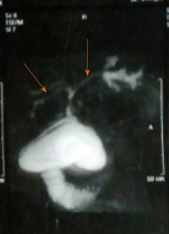
Figure 2 Magnetic resonance cholangiopancreatography of a patient with inflammatory bowel disease and primary sclerosing cholangitis showing dilatation of the intra- and extra-hepatic biliary tree with multiple strictures (arrows).
Figure 3 Histopathological findings of a liver biopsy specimen from a patient with inflammatory bowel disease showing the features of primary sclerosing cholangitis.
A: Diffuse bile ductular proliferation "Onion skin" fibrosis around affected ducts, concentric collagen fibre deposition; B: Same findings by Masson's trichrome stain which imparts a blue color to type 1 collagen against a red background of hepatocytes; C: Aggression of inflammatory cells towards the biliary epithelium and the portal area is markedly expanded with lymphoplasmacytic inflammatory cellular infiltrate (all photos H&E, Original magnification × 400).
- Citation: El-Shabrawi MH, Tarek S, Abou-Zekri M, Meshaal S, Enayet A, Mogahed EA. Hepatobiliary manifestations in children with inflammatory bowel disease: A single-center experience in a low/middle income country. World J Gastrointest Pharmacol Ther 2020; 11(3): 48-58
- URL: https://www.wjgnet.com/2150-5349/full/v11/i3/48.htm
- DOI: https://dx.doi.org/10.4292/wjgpt.v11.i3.48


