Published online Sep 28, 2023. doi: 10.4329/wjr.v15.i9.274
Peer-review started: June 30, 2023
First decision: August 24, 2023
Revised: September 4, 2023
Accepted: September 22, 2023
Article in press: September 22, 2023
Published online: September 28, 2023
Processing time: 88 Days and 17.8 Hours
Primary central nervous system lymphoma (PCNSL) is a rare malignant tumor originating from the lymphatic hematopoietic system. It exhibits unique imaging manifestations due to its biological characteristics.
Magnetic resonance imaging (MRI) with diffusion-weighted imaging (DWI), perfusion-weighted imaging (PWI), and magnetic resonance spectroscopy was performed. The imaging findings showed multiple space-occupying lesions with low signal on T1-weighted imaging, uniform high signal on T2-weighted imaging, and obvious enhancement on contrast-enhanced scans. DWI revealed diffusion restriction, PWI demonstrated hypoperfusion, and spectroscopy showed elevated choline peak and decreased N-acetylaspartic acid. The patient's condition significantly improved after hormone shock therapy.
This case highlights the distinctive imaging features of PCNSL and their importance in accurate diagnosis and management.
Core Tip: Primary central nervous system lymphoma (PCNSL) is a rare tumor of the central nervous system with distinctive imaging features. This case report highlights the imaging manifestations of multiple PCNSL lesions using diffusion-weighted imaging, perfusion-weighted imaging, and magnetic resonance imaging. Accurate diagnosis is crucial for appropriate management. Further research and larger studies are needed to enhance the understanding and diagnostic accuracy of PCNSL.
- Citation: Liu LH, Zhang HW, Zhang HB, Liu XL, Deng HZ, Lin F, Huang B. Distinctive magnetic resonance imaging features in primary central nervous system lymphoma: A case report. World J Radiol 2023; 15(9): 274-280
- URL: https://www.wjgnet.com/1949-8470/full/v15/i9/274.htm
- DOI: https://dx.doi.org/10.4329/wjr.v15.i9.274
Primary central nervous system lymphoma (PCNSL) is an exceptionally rare and aggressive malignancy originating within the central nervous system. Despite its infrequency, PCNSL presents unique diagnostic and therapeutic challenges, rendering it a subject of profound clinical importance[1]. In recent years, advanced imaging techniques, including diffusion-weighted imaging (DWI), perfusion-weighted imaging (PWI), and magnetic resonance (MR) spectroscopy, have greatly enhanced our ability to comprehend the disease and its distinctive manifestations[2].
While previous studies have provided valuable insights into the broader understanding of PCNSL, there is a critical gap in characterizing the imaging features on an individual case level[3]. This study endeavors to bridge this gap by conducting an in-depth examination of DWI, PWI, and MR spectroscopy findings in a single PCNSL case. By scrutinizing these imaging modalities within the unique context of this individual case, we aim to contribute to the comprehensive understanding of PCNSL's imaging features and their potential clinical implications[4].
The primary research problem addressed in this study is to delineate the specific magnetic resonance imaging (MRI) imaging manifestations of PCNSL within the scope of a single case analysis and to comprehend their diagnostic and clinical significance. To accomplish this, we have conducted a detailed analysis of DWI, PWI, and MR spectroscopy findings in the context of this singular PCNSL case.
A 79-year-old female patient with a previously unremarkable medical history presented with a sudden onset of unexplained dizziness accompanied by projectile vomiting, characterized by the ejection of gastric contents. She also reported a sense of heaviness and weakness in her limbs.
Initial evaluation at another medical facility revealed the presence of multiple intracranial space-occupying lesions. These lesions were detected through a computed tomography scan, which indicated the involvement of the cerebellar hemisphere, corpus callosum's splenium, and the left parietal lobe.
No special notes.
No special notes.
Physical examination revealed no abnormalities.
The platelet specific volume was slightly elevated. The monocyte count was mildly elevated. The bacterial content in the urine test increased [4225.40, reference value: 0-4000 (/μL)].
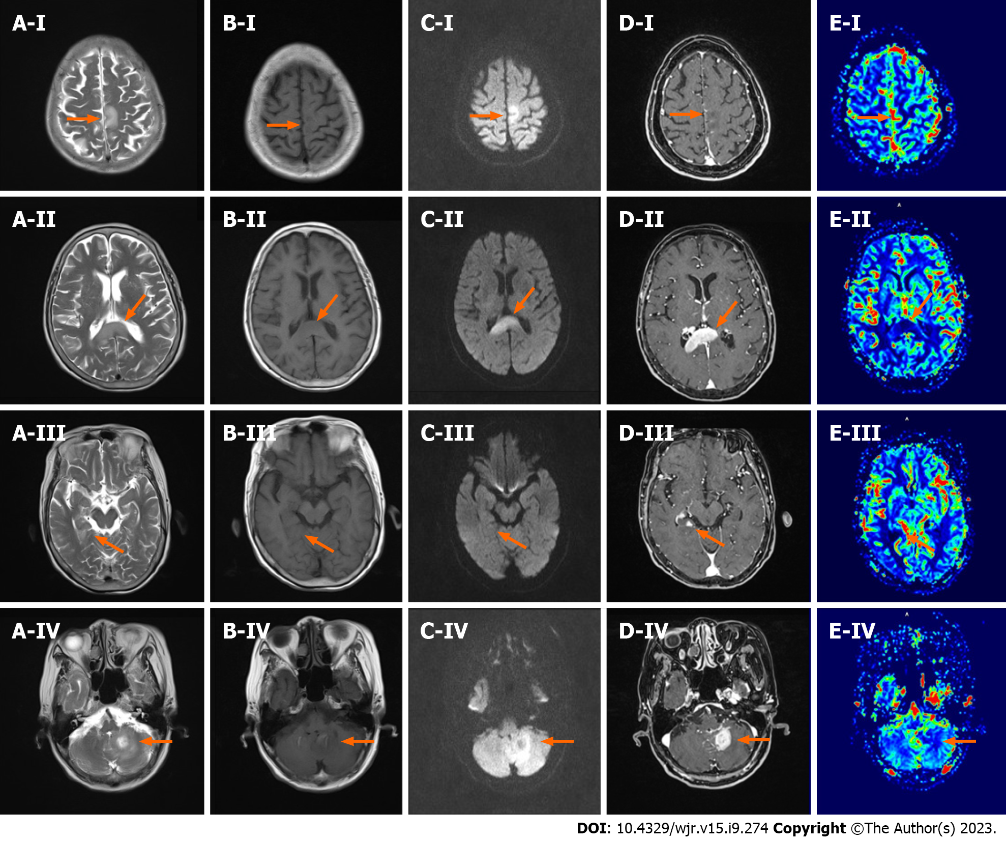
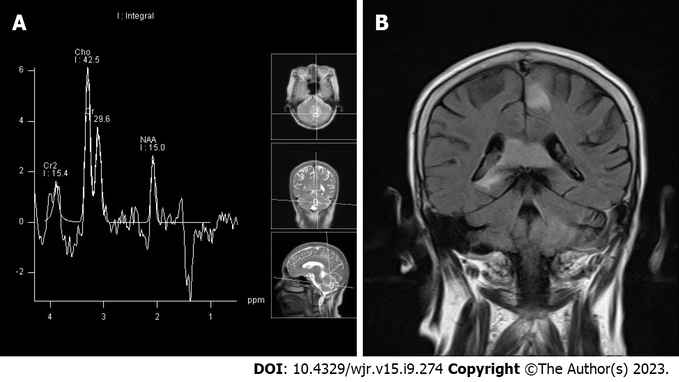
Upon admission, the patient underwent a comprehensive evaluation, including MRI with DWI, PWI, and MR spectroscopy (MRS). The MRI results confirmed the presence of multiple space-occupying lesions, characterized by low signal intensity on T1-weighted imaging, uniform high signal intensity on T2-weighted imaging, and prominent enhancement on contrast-enhanced scans (Figure 1). DWI further revealed diffusion restriction, while PWI demonstrated hypoperfusion in all the identified lesions. Additionally, spectroscopy (MRS) depicted an elevated choline peak and decreased N-acetylaspartic acid. Notably, MRS also revealed the presence of a Lip peak within the lesion (Figure 2). These combined imaging features strongly suggested the possibility of PCNSL.
Pathological examination of the intracranial lesion confirmed the presence of an aggressive B-cell lymphoma. Immunohistochemical analysis demonstrated the following profile: CD21 (-), CD10 (-), CD20 (+), CD3 (background T-cells +), CD30 (-), PAX-5 (+), Bcl-6 (+), MUM-1 (+), Ki-67 (approximately 80%), CD5 (-), CD23 (-), Bcl-2 (-), CyclinD1 (-), CD79A (+), C-MYC (approximately 30%+), P53 (10%+ with varying intensity), GFAP (glial cells +). Importantly, Epstein-Barr virus-encoded small RNA (EBER) was not detected. These findings confirmed the diagnosis of diffuse large B-cell lymphoma of the non-germinal center B-cell (non-GCB) subtype, supporting the diagnosis of PCNSL.
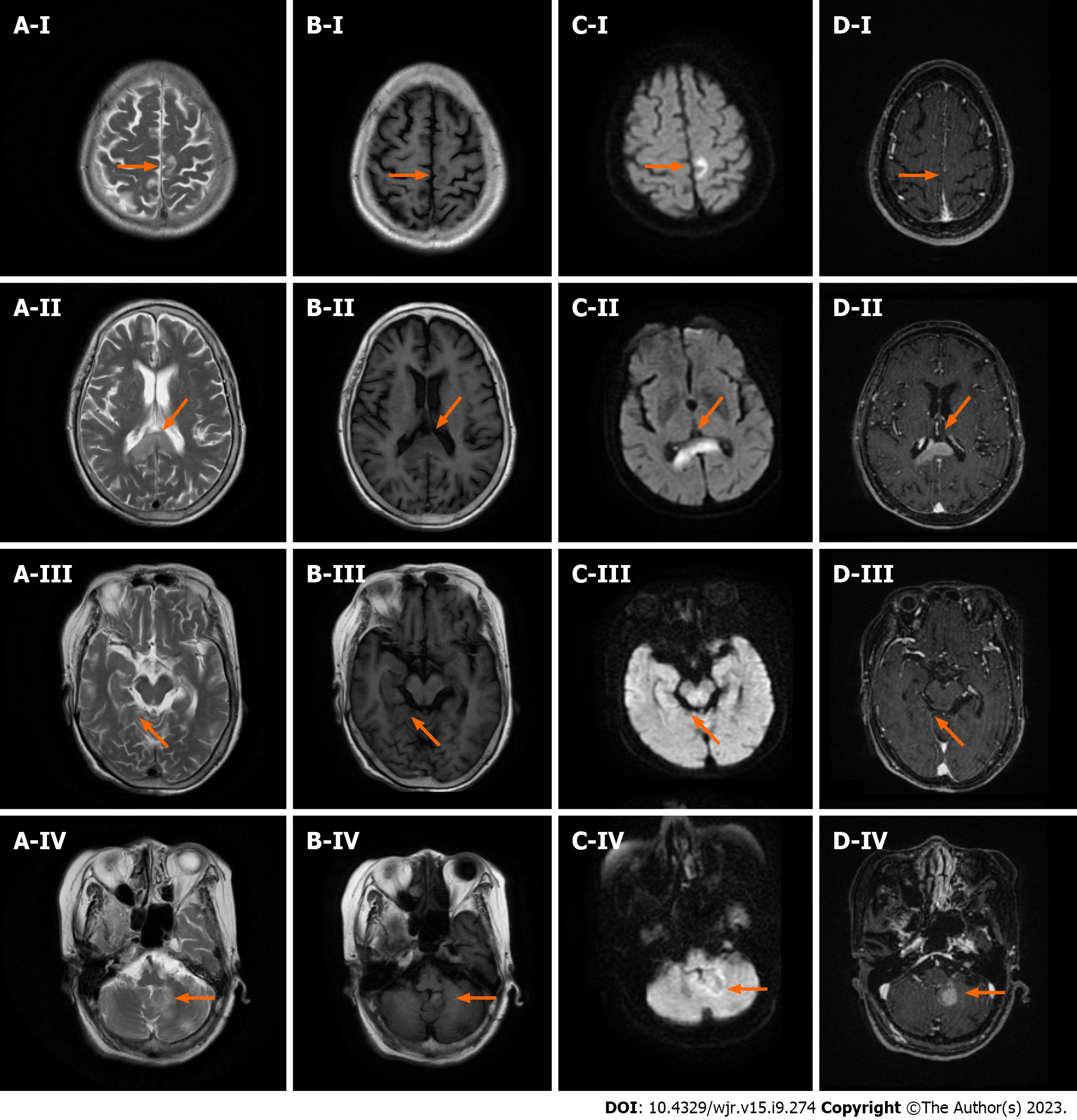
In response to the initial presentation and MRI findings, the patient underwent a diagnostic trial of steroid therapy. This treatment led to a significant improvement in the patient's neurological condition, although it was accompanied by the emergence of certain neuropsychiatric symptoms. Subsequent administration of medications, including lorazepam and olanzapine, resulted in a notable improvement in the patient's neuropsychiatric symptoms. Follow-up MRI examinations indicated a reduction in enhancement in the lesions located in the hippocampus and left parietal lobe, with the other lesions remaining stable or showing slight reductions (Figure 3).
The patient subsequently received standardized lymphoma immunotherapy and chemotherapy regimens, and her prognosis remains favorable.
There is no lymphoid tissue present within the central nervous system, making brain lymphomas relatively rare. Currently, the origin of intracerebral lymphoma can be attributed to the following factors: Firstly, reactive lymphocytes enter the brain tissue during intracerebral infection and undergo malignant transformation through various mechanisms. Secondly, activated peripheral lymphocytes transform into tumor cells and migrate to the brain through the bloodstream, resulting in tumors primarily located around the ventricles, basal ganglia, and frontoparietal lobes. Thirdly, undifferentiated pluripotent stem cells surrounding blood vessels in the brain may serve as the source of intracerebral lymphoma. Histologically, intracerebral lymphomas exhibit predominant sleeve-shaped growth, infiltrating the surrounding brain parenchyma, and demonstrating multicentric growth within the tumor[5].
PCNSL typically presents as supratentorial lesions, with predilection sites in the cerebral hemisphere, corpus callosum, basal ganglia, and thalamus[6]. The imaging findings of PCNSL are closely related to its pathological features. In this case, the lesions were confined to the brain tissue, involving both supratentorial and infratentorial areas, which is relatively rare[7].
On conventional MRI, PCNSL shows iso-hypointensity on T1-weighted imaging and iso-hypointensity on T2-weighted imaging[8]. DWI demonstrates diffusion restriction due to densely arranged tumor cells. According to Lin et al[9], their study suggests that combining DWI ADC value with T1WI enhanced scan can aid in the differentiation of glioblastoma from PCNSL. PWI reveals hypoperfusion, reflecting the hypo-vascular nature of PCNSL[10]. Contrast-enhanced scans show uniform enhancement when the tumor invades adjacent brain parenchyma and disrupts the blood-brain barrier (BBB). In our previous study, we compared high-grade glioma (HGG) with lymphoma using dynamic contrast-enhanced (DCE) imaging, and found that lymphoma has more obvious damage to the BBB, resulting in transfer constant(Ktrans) values even higher than HGG[11]. The characteristic imaging features, such as the "fist sign," "sharp horn sign," and "butterfly wing sign,"(usually occurs in the corpus callosum) are associated with the tumor's angiophilic growth.
MRS plays a crucial role in the evaluation of PCNSL. Elevated choline peak, decreased N-acetylaspartic acid, and a towering lipid peak are commonly observed[12]. The towering lipid peak is highly specific for the diagnosis of PCNSL, attributed to the accelerated turnover of lymphocytes and macrophages. PCNSL should be differentiated from demyelinating diseases, as their imaging findings may resemble each other[13]. Hormone shock therapy can provide symptomatic relief in both PCNSL and demyelinating diseases, but recovery of symptoms after hormone therapy withdrawal is indicative of PCNSL.
In addition to our contributions to the understanding of PCNSL, it is essential to recognize the limitations of our study. The utilization of a single-case design inherently limits the generalizability of our findings to a broader population of PCNSL patients. Future research should strive to replicate these findings in larger cohorts to establish their broader applicability. Furthermore, our study primarily focuses on the imaging aspects of PCNSL, and future investigations could explore the correlation between imaging features and specific clinical outcomes. These considerations highlight both the strengths and the areas for improvement in our research.
This single-case analysis of PCNSL has shed light on the distinctive MRI imaging features of this rare malignancy. By employing advanced techniques such as DWI, PWI, and MR spectroscopy, we have provided a comprehensive characterization of PCNSL's imaging manifestations. However, it is important to acknowledge the limitations of this study. The use of a single-case design restricts the generalizability of our findings to a broader population. Future research should aim to replicate these findings in larger cohorts of PCNSL patients to establish their broader applicability. Additionally, our study focused on the imaging aspects, and future investigations could explore the correlation between imaging features and specific clinical outcomes. Despite these limitations, our study contributes valuable insights into the unique imaging features of PCNSL and serves as a foundation for further research in this area.
Provenance and peer review: Unsolicited article; Externally peer reviewed.
Peer-review model: Single blind
Specialty type: Radiology, nuclear medicine and medical imaging
Country/Territory of origin: China
Peer-review report’s scientific quality classification
Grade A (Excellent): 0
Grade B (Very good): B
Grade C (Good): 0
Grade D (Fair): D
Grade E (Poor): 0
P-Reviewer: Mahmoud MZ, Saudi Arabia S-Editor: Liu JH L-Editor: A P-Editor: Liu JH
| 1. | Krebs S, Barasch JG, Young RJ, Grommes C, Schöder H. Positron emission tomography and magnetic resonance imaging in primary central nervous system lymphoma-a narrative review. Ann Lymphoma. 2021;5. [RCA] [PubMed] [DOI] [Full Text] [Full Text (PDF)] [Cited by in Crossref: 4] [Cited by in RCA: 18] [Article Influence: 3.6] [Reference Citation Analysis (0)] |
| 2. | Schaff LR, Grommes C. Primary central nervous system lymphoma. Blood. 2022;140:971-979. [RCA] [PubMed] [DOI] [Full Text] [Cited by in Crossref: 33] [Cited by in RCA: 146] [Article Influence: 36.5] [Reference Citation Analysis (0)] |
| 3. | Deng B, Dai Y, Wang Q, Yang J, Chen X, Liu TT, Liu J. The clinical analysis of new-onset status epilepticus. Epilepsia Open. 2022;7:771-780. [RCA] [PubMed] [DOI] [Full Text] [Full Text (PDF)] [Cited by in Crossref: 1] [Cited by in RCA: 6] [Article Influence: 1.5] [Reference Citation Analysis (0)] |
| 4. | Han Y, Wang ZJ, Li WH, Yang Y, Zhang J, Yang XB, Zuo L, Xiao G, Wang SZ, Yan LF, Cui GB. Differentiation Between Primary Central Nervous System Lymphoma and Atypical Glioblastoma Based on MRI Morphological Feature and Signal Intensity Ratio: A Retrospective Multicenter Study. Front Oncol. 2022;12:811197. [RCA] [PubMed] [DOI] [Full Text] [Full Text (PDF)] [Cited by in Crossref: 1] [Cited by in RCA: 13] [Article Influence: 3.3] [Reference Citation Analysis (0)] |
| 5. | Barajas RF, Politi LS, Anzalone N, Schöder H, Fox CP, Boxerman JL, Kaufmann TJ, Quarles CC, Ellingson BM, Auer D, Andronesi OC, Ferreri AJM, Mrugala MM, Grommes C, Neuwelt EA, Ambady P, Rubenstein JL, Illerhaus G, Nagane M, Batchelor TT, Hu LS. Consensus recommendations for MRI and PET imaging of primary central nervous system lymphoma: guideline statement from the International Primary CNS Lymphoma Collaborative Group (IPCG). Neuro Oncol. 2021;23:1056-1071. [RCA] [PubMed] [DOI] [Full Text] [Full Text (PDF)] [Cited by in Crossref: 18] [Cited by in RCA: 115] [Article Influence: 23.0] [Reference Citation Analysis (0)] |
| 6. | Weller M. The vanishing role of whole brain radiotherapy for primary central nervous system lymphoma. Neuro Oncol. 2014;16:1035-1036. [RCA] [PubMed] [DOI] [Full Text] [Cited by in Crossref: 9] [Cited by in RCA: 10] [Article Influence: 0.8] [Reference Citation Analysis (0)] |
| 7. | Bhattacharjee R, Gupta M, Singh T, Sharma S, Khanna G, Parvaze SP, Patir R, Vaishya S, Ahlawat S, Singh A, Gupta RK. Role of intra-tumoral vasculature imaging features on susceptibility weighted imaging in differentiating primary central nervous system lymphoma from glioblastoma: a multiparametric comparison with pathological validation. Neuroradiology. 2022;64:1801-1818. [RCA] [PubMed] [DOI] [Full Text] [Cited by in RCA: 6] [Reference Citation Analysis (0)] |
| 8. | Toh CH, Wei KC, Chang CN, Ng SH, Wong HF. Differentiation of primary central nervous system lymphomas and glioblastomas: comparisons of diagnostic performance of dynamic susceptibility contrast-enhanced perfusion MR imaging without and with contrast-leakage correction. AJNR Am J Neuroradiol. 2013;34:1145-1149. [RCA] [PubMed] [DOI] [Full Text] [Cited by in Crossref: 77] [Cited by in RCA: 93] [Article Influence: 7.2] [Reference Citation Analysis (0)] |
| 9. | Lin X, Lee M, Buck O, Woo KM, Zhang Z, Hatzoglou V, Omuro A, Arevalo-Perez J, Thomas AA, Huse J, Peck K, Holodny AI, Young RJ. Diagnostic Accuracy of T1-Weighted Dynamic Contrast-Enhanced-MRI and DWI-ADC for Differentiation of Glioblastoma and Primary CNS Lymphoma. AJNR Am J Neuroradiol. 2017;38:485-491. [RCA] [PubMed] [DOI] [Full Text] [Cited by in Crossref: 53] [Cited by in RCA: 69] [Article Influence: 6.9] [Reference Citation Analysis (0)] |
| 10. | Kang KM, Choi SH, Chul-Kee P, Kim TM, Park SH, Lee JH, Lee ST, Hwang I, Yoo RE, Yun TJ, Kim JH, Sohn CH. Differentiation between glioblastoma and primary CNS lymphoma: application of DCE-MRI parameters based on arterial input function obtained from DSC-MRI. Eur Radiol. 2021;31:9098-9109. [RCA] [PubMed] [DOI] [Full Text] [Cited by in Crossref: 2] [Cited by in RCA: 15] [Article Influence: 3.0] [Reference Citation Analysis (0)] |
| 11. | Zhang HW, Lyu GW, He WJ, Lei Y, Lin F, Feng YN, Wang MZ. Differential diagnosis of central lymphoma and high-grade glioma: dynamic contrast-enhanced histogram. Acta Radiol. 2020;61:1221-1227. [RCA] [PubMed] [DOI] [Full Text] [Cited by in Crossref: 4] [Cited by in RCA: 12] [Article Influence: 2.0] [Reference Citation Analysis (0)] |
| 12. | Du X, He Y, Lin W. Diagnostic Accuracy of the Diffusion-Weighted Imaging Method Used in Association With the Apparent Diffusion Coefficient for Differentiating Between Primary Central Nervous System Lymphoma and High-Grade Glioma: Systematic Review and Meta-Analysis. Front Neurol. 2022;13:882334. [RCA] [PubMed] [DOI] [Full Text] [Full Text (PDF)] [Cited by in Crossref: 1] [Cited by in RCA: 8] [Article Influence: 2.0] [Reference Citation Analysis (0)] |
| 13. | Miyajima T, Ohigashi H, Yaguchi H, Teshima T. Neurolymphomatosis in Intravascular Large B-cell Lymphoma. Intern Med. 2023;62:1381-1382. [RCA] [PubMed] [DOI] [Full Text] [Reference Citation Analysis (0)] |


