©The Author(s) 2017.
World J Radiol. Mar 28, 2017; 9(3): 143-147
Published online Mar 28, 2017. doi: 10.4329/wjr.v9.i3.143
Published online Mar 28, 2017. doi: 10.4329/wjr.v9.i3.143
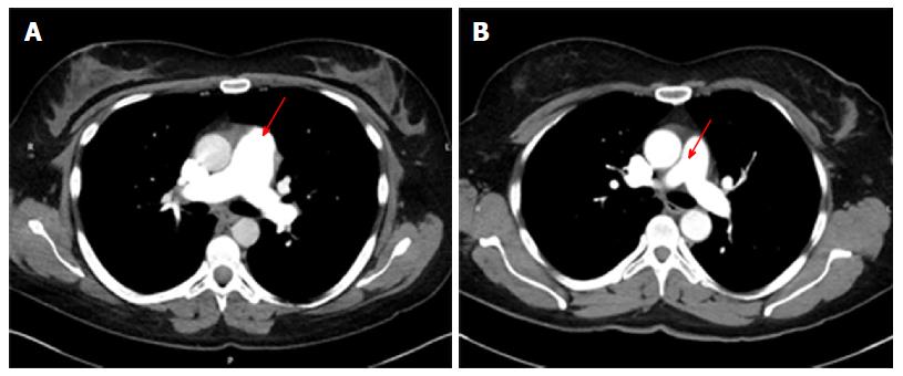
Figure 1 Sample axial image slices showing the good opacification in the main pulmonary artery with intravenous contrast in the two groups (red arrows).
A: 60 mL; B: 75 mL.
- Citation: Chen M, Mattar G, Abdulkarim JA. Computed tomography pulmonary angiography using a 20% reduction in contrast medium dose delivered in a multiphasic injection. World J Radiol 2017; 9(3): 143-147
- URL: https://www.wjgnet.com/1949-8470/full/v9/i3/143.htm
- DOI: https://dx.doi.org/10.4329/wjr.v9.i3.143


