Published online Mar 28, 2017. doi: 10.4329/wjr.v9.i3.134
Peer-review started: November 2, 2016
First decision: December 15, 2016
Revised: December 22, 2016
Accepted: January 11, 2017
Article in press: January 14, 2017
Published online: March 28, 2017
Processing time: 143 Days and 10.5 Hours
To assess the relationship using multimodality imaging between intermediary citrate/choline metabolism as seen on proton magnetic resonance spectroscopic imaging (1H-MRSI) and glycolysis as observed on 18F-fluorodeoxyglucose positron emission tomography/computed tomography (18F-FDG-PET/CT) in prostate cancer (PCa) patients.
The study included 22 patients with local PCa who were referred for endorectal magnetic resonance imaging/1H-MRSI (April 2002 to July 2007) and 18F-FDG-PET/CT and then underwent prostatectomy as primary or salvage treatment. Whole-mount step-section pathology was used as the standard of reference. We assessed the relationships between PET parameters [standardized uptake value (SUVmax and SUVmean)] and MRSI parameters [choline + creatine/citrate (CC/Cmax and CC/Cmean) and total number of suspicious voxels] using spearman’s rank correlation, and the relationships of PET and 1H-MRSI index lesion parameters to surgical Gleason score.
Abnormal intermediary metabolism on 1H-MRSI was present in 21/22 patients, while abnormal glycolysis on 18F-FDG-PET/CT was detected in only 3/22 patients. Specifically, index tumor localization rates were 0.95 (95%CI: 0.77-1.00) for 1H-MRSI and 0.14 (95%CI: 0.03-0.35) for 18F-FDG-PET/CT. Spearman rank correlations indicated little relationship (ρ = -0.36-0.28) between 1H-MRSI parameters and 18F-FDG-PET/CT parameters. Both the total number of suspicious voxels (ρ = 0.55, P = 0.0099) and the SUVmax (ρ = 0.46, P = 0.0366) correlated weakly with the Gleason score. No significant relationship was found between the CC/Cmax, CC/Cmean or SUVmean and the Gleason score (P = 0.15-0.79).
The concentration of intermediary metabolites detected by 1H MRSI and glycolytic flux measured 18F-FDG PET show little correlation. Furthermore, only few tumors were FDG avid on PET, possibly because increased glycolysis represents a late and rather ominous event in the progression of PCa.
Core tip: Although metabolic imaging is increasingly utilized in prostate cancer (PCa), the mechanisms leading to cancer-related metabolic rearrangements and consequent imaging findings remain poorly understood. This study compared two modalities utilizing distinct metabolic pathways, proton magnetic resonance spectroscopic imaging (1H-MRSI) and 18F-fluorodeoxyglucose positron emission tomography/computed tomography (18F-FDG-PET/CT), in local PCa. Abnormal intermediary metabolism on 1H-MRSI was present in 21/22 patients, while abnormal glycolysis on 18F-FDG-PET/CT was detected in only 3/22 patients. This study provides an insight why metabolic PET agents promising for detection of PCa target intermediary metabolism. On the other hand, elevated glycolysis may have ominous prognostic implications in PCa.
- Citation: Shukla-Dave A, Wassberg C, Pucar D, Schöder H, Goldman DA, Mazaheri Y, Reuter VE, Eastham J, Scardino PT, Hricak H. Multimodality imaging using proton magnetic resonance spectroscopic imaging and 18F-fluorodeoxyglucose-positron emission tomography in local prostate cancer. World J Radiol 2017; 9(3): 134-142
- URL: https://www.wjgnet.com/1949-8470/full/v9/i3/134.htm
- DOI: https://dx.doi.org/10.4329/wjr.v9.i3.134
Multimodality imaging is performed in patients with cancer to understand metabolism, however the mechanisms leading to metabolic rearrangements remains poorly understood. By decoding the connections between cancer signaling and metabolism, it may be possible to better understand the clinical implications of imaging findings and develop new imaging strategies.
In patients with prostate cancer (PCa), two key functional imaging modalities, proton magnetic resonance spectroscopic imaging (1H-MRSI) and 18F-fluorodeoxyglucose positron emission tomography/computed tomography (18F-FDG-PET/CT), are used to identify cancer-induced changes in cellular metabolism[1-3]. On 1H-MRSI, decreased levels of citrate (a Krebs cycle and fatty acid synthesis intermediate) and polyamines (amino acid metabolism intermediates) and elevated choline (a precursor of membrane synthesis) are a signature of PCa[2,4-7]. On 18F-FDG-PET, increased glucose uptake by glucose transporters and glucose phosphorylation to glucose-6-phosphate by hexokinase are used for identifying PCa and other cancers[3,8]. The indications for 1H-MRSI and 18F-FDG-PET/CT examinations in patients with PCa are very different[9,10]. 1H-MRSI adds incremental value to standard prostate magnetic resonance imaging (MRI) in the detection of primary or recurrent loco-regional PCa and in the evaluation of its aggressiveness (Gleason Grade)[9-14]. 1H-MRSI can also be used prior to treatment to predict biochemical relapse of PCa after radical prostatectomy or the presence of insignificant PCa[14-17]. In contrast, 18F-FDG-PET/CT is not recommended for the detection and initial evaluation of PCa or detection of early recurrence. Various other PET agents, such as 18F- or 11C-labeled choline or acetate, several various amino acids, and agents binding to the transmembrane PSMA molecule are available for this purpose[18-24]. 18F-FDG plays a role in the characterization of advanced metastatic PCa[8,25]. The clinical significance of the current study lies in exploring the use of multimodality imaging in local PCa. In this study, we wanted to explore the relationship using multimodality imaging between concentrations of intermediary metabolites citrate and choline, measured by 1H-MRSI, and glycolysis as noted on 18F-FDG PET/CT in local PCa.
Our study was compliant with the Health Insurance Portability and Accountability Act. Patient data were collected and handled in accordance with institutional and federal guidelines. Twenty-two patients who were referred from the Urology department for endorectal MRI/MRSI examinations (April 2002 to July 2007) and 18F-FDG-PET/CT and then underwent prostatectomy as primary or salvage treatment were included in the study. Whole-mount step-section pathology of the surgical specimen was available for all the patients. Of the 22 patients, 11 were imaged before treatment while 11 were imaged after external beam radiation therapy. The institutional review board approved our retrospective review of multimodality imaging using MRI/MRSI and 18F-FDG-PET/CT studies, pathology data (from surgical pathology), and clinical follow-up data and waived the informed consent requirement. The mean time between the MRSI and PET exams was 11 ± 37 d (± SD).
Data were acquired on a 1.5-Tesla GE Signa Horizon scanner. MRI was done using a pelvic phased-array coil and an expandable endorectal coil; T1- and T2-weighted spin-echo MR images were obtained using a previously described standard prostate imaging protocol (total time, approximately 30 min)[4]. MR image acquisition was followed by a standard MRSI protocol with point-resolved spectroscopic voxel excitation and water and lipid suppression (total time, 17 min) in a voxel array and the SI dimension zero filled to 16 slices (3-mm resolution) with a voxel size of 0.12 to 0.16 cm3[4]. MRSI data were overlaid on the corresponding T2-weighted images, including the raw spectra and the metabolic ratio [choline + creatine to citrate (CC/C)][4]. Tumors were identified by dedicated radiologists with > 5 years of experience in prostate imaging.
An MRI physicist with > 10 years of experience in prostate MRSI retrospectively interpreted the 1H-MRSI studies using established metabolic criteria for the evaluation of PCa in the peripheral and transition zones[6,14,26,27]. The physicist, who was blinded to clinical data and surgical pathology, recorded the location and total number of suspicious voxels (tumor volume estimation), maximum (max) CC/C, and mean CC/C for the index lesion[6,14,26,27].
Details of the 18F-FDG-PET/CT imaging procedure have been described previously[28]. Briefly, a low-dose CT scan (120-140 kV, approximately 80 mA), which is used for attenuation correction of PET emission images as well as for anatomic localization of PET abnormalities, was acquired first. This was followed by acquisition of PET emission images of the lower pelvis including of the prostate for 5 min per bed position. Images were reconstructed using iterative algorithms with average slice thickness of 3 mm and a 128 × 128 matrix size. Patients were scanned in the supine position. Before the examination, patients fasted for at least 6 h, but liberal intake of water was allowed. Patients were injected intravenously with 444-555 MBq of 18F-FDG and a PET/CT scan started after an uptake period of approximately 60 min. Plasma glucose level was < 200 mg at the time of imaging in all patients.
All 18F-FDG-PET/CT data were available for retrospective review on a standard clinical workstation (PACS with Advance Work Station extension; General Electric). One board-certified radiologist/nuclear medicine physician, who had > 10 years of experience in PET and > 5 years of experience in prostate imaging, reviewed the 18F-FDG-PET/CT studies. PET images were analyzed in three orthogonal planes (transaxial, coronal, sagittal) both visually and quantitatively. For quantitative PET analysis, maximum standardized uptake value (SUVmax) and average SUV (SUVmean) of the index lesion were determined using a Volume of Interest with 40% threshold of SUVmax. All SUVs were normalized to body weight.
Whole-mount transverse serial sections of the prostate were prepared as described previously[29]. The distal 5-mm portion of the apex was amputated and coned. The remainder of the gland was serially sectioned from the apex to the base at 3-4-mm intervals and submitted in its entirety for paraffin-embedded whole mounts. Cancer foci were outlined in ink on whole-mount, apical, and seminal vesicle sections and photographed. The photographs constituted the tumor maps. The primary and secondary Gleason grades as well as the pathologic tumor node stage were also determined. The index lesion was identified in all cases as the tumor with the largest volume. Tissue sections stained with H and E were examined by one uro-pathologist blinded to imaging and clinical data.
The matching of imaging and pathology for the index lesion was performed in consensus by two dually-boarded radiologists/nuclear medicine physicians with > 5 years and > 15 years of experience in prostate imaging. The histopathologic axial step sections were visually matched with corresponding axial T2-weighted transverse MR images with superimposed MRSI data and fused 18F-FDG PET/CT data with a precision of ± 1 slice based on established anatomic landmarks[12]. Because the spectroscopic data were acquired in the same position and with the same gradients as the imaging data, registration of the spectroscopic data with the T2-weighted images was automatic, and the spectroscopic data could be compared with the most closely corresponding histopathologic step section.
Clinical and pathological characteristics were described using medians and ranges for continuous variables and frequencies and percents or proportions for categorical variables. Gleason grades were summed into Gleason scores of 6, 7, 8 or 9.
The localization rates of 18F-FDG-PET/CT and 1H-MRSI were calculated along with exact 95% confidence intervals. The relationships between PET parameters (SUVmax and SUVmean) and MRSI parameters (CC/Cmax, CC/Cmean, Total # Voxels) were assessed using Spearman’s rank correlation and graphically displayed with scatter plots and 95% confidence bands. Additionally, the relationships of PET and MRSI parameters to surgical Gleason score were assessed with Spearman’s rank correlation and, given the Gleason score’s ordinal nature, graphically illustrated with box plots. P-values less than 0.05 were considered statistically significant. All analyses were done using SAS 9.4 (The SAS Institute, Cary, NC).
Patient characteristics are summarized in Table 1. The patients had a median age of 58 years (range: 47-70 years) and median PSA of 4.81 ng/mL (range: 0.11-96.53 ng/mL).
| n (%) | ||
| Clinical stage1 | T1c | 10 (45.5) |
| T2a | 4 (18.2) | |
| T2b | 4 (18.2) | |
| T2c | 1 (4.5) | |
| T3 | 1 (4.5) | |
| T3a | 2 (9.1) | |
| Biopsy Gleason score | 0 + 0 | 1 (4.5) |
| 3 + 3 | 3 (13.6) | |
| 3 + 4 | 5 (22.7) | |
| 4 + 3 | 5 (22.7) | |
| 4 + 4 | 4 (18.2) | |
| 4 + 5 | 4 (18.2) | |
| Pathology stage | pT2a | 3 (13.6) |
| pT2b | 5 (22.7) | |
| pT3a | 7 (31.8) | |
| pT3b | 6 (27.3) | |
| pT4 | 1 (4.5) | |
| Pathology Gleason score | 3 + 3 | 1 (4.5) |
| 3 + 4 | 8 (36.4) | |
| 4 + 3 | 4 (18.2) | |
| 4 + 4 | 4 (18.2) | |
| 4 + 5 | 3 (13.6) | |
| 5 + 4 | 1 (4.5) | |
| Not Graded2 | 1 (4.5) | |
| Prior radiation treatment | EBRT | 11 (50) |
| Untreated | 11 (50) | |
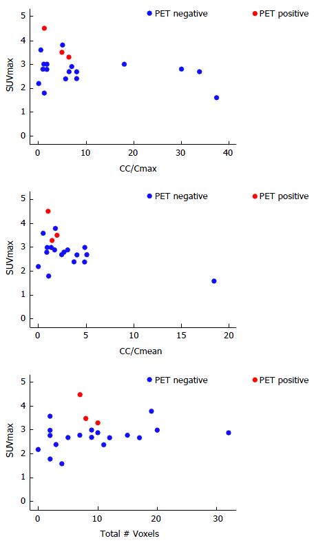
Index tumor localization rates were 0.95 (95%CI: 0.77-1.00) for 1H-MRSI and 0.14 (95%CI: 0.03-0.35) for 18F-FDG-PET/CT, with 21 out of 22 index tumors found on pathology identified on 1H-MRSI and only 3 of those 21 index lesions identified on 18F-FDG-PET/CT. Figure 1 shows 1H-MRSI, 18F-FDG-PET/CT and whole-mount step-section pathology from a patient in whom the tumor seen at pathology was observed by multimodality imaging. Figure 2 shows 1H-MRSI, 18F-FDG-PET/CT and whole-mount step-section pathology from a patient in whom the tumor seen at pathology was observed by 1H-MRSI only. In the 3 patients with positive PET findings, the total tumor volumes measured by PET were 10.9, 11.1 and 10.4 cc and the SUVmax values were 3.3, 3.5 and 4.5. On 1H-MRSI in the 21 positive patients, CC/Cmax (median 6.4, range: 0.5-37.4), CC/Cmean (median 2.0, range: 0.5-18.5) and number of suspicious voxels (median 9.0, range 2-32) showed more profound alterations for all patients. Both the scatter plots (Figure 3 and Table 2) and the Spearman rank correlations indicated little relationship between 1H-MRSI parameters and 18F-FDG-PET/CT. Spearman’s ρ ranged between -0.362 and 0.28 (P-values range: 0.10-0.66). No clear pattern of association was detected in the graphs.
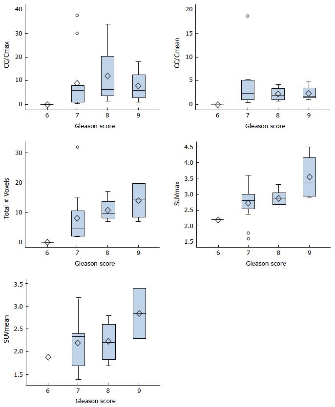
Gleason scores ranged from 6 (8/22, 36%) to 9 (4/22, 18.1%) with one patient lacking a score due to treatment effect. This patient was excluded from further analysis. Both the total number of voxels (ρ = 0.55, P = 0.0099) and the SUVmax (ρ = 0.46, P = 0.0366) correlated with the Gleason score. No significant relationship was found between the CC/Cmax, CC/Cmean or SUVmean and the Gleason score (P = 0.15-0.79, Table 3). The box plots demonstrate an upward trend for total number of voxels and SUVmax with each subsequent Gleason score (Figure 4).
Multimodality imaging in PCa detection on 1H-MRSI is based on the detection of decreased citrate and polyamines with elevated choline[2,4]. This is reflected in the high CC/Cmax (median 9.0) and CC/Cmean (median: 2.0) for the prostate index lesions. On 18F-FDG-PET/CT, PCa is identified based on increased glucose uptake by glucose transporters (GLUT) and glucose phosphorylation to glucose-6-phosphate by hexokinase[3,8]. The present study adds to the literature for patients with local or loco-regional primary or recurrent PCa and shows that the citrate decrease in PCa was both much more frequent and pronounced than was the elevation in 18F-FDG uptake. This is in-line with the known low sensitivity of 18F-FDG-PET/CT for detecting localized primary PCa. Further research is needed to develop a clearer understanding of the underlying genomic and metabolic mechanisms and to confirm whether metabolic alterations progress stepwise from early abnormalities in citrate metabolism to late abnormalities in glucose metabolism. Since our patients were imaged at only one time point, the data appear consistent with this understanding. Improved understanding of PCa metabolism could help in determining the most appropriate imaging modalities (including imaging with radiotracers) for different clinical stages of PCa and possibly also in identifying and monitoring novel targeted therapies.
For instance, according to the “bioenergetic theory of prostate malignancy”[1,17], the normal prostate produces and secretes an enormous amount of citrate; this is achieved by zinc-induced inhibition of m-aconitase, a Krebs cycle enzyme that converts citrate to isocitrate. With this truncated Krebs cycle, the normal prostate sacrifices ATP production for citrate secretion[17]. Conversely, in PCa, down-regulation of multiple membrane zinc transporters and zinc decline lead to activation of the full Krebs cycle, oxidation and a consequent decrease in intracellular and secreted citrate and increase in ATP production supporting malignancy[30-32]. However, the decline of citrate in PCa could also be related to its conversion to AcCoA by cytosolic ATP citrate lyase (ACLY) and subsequent utilization for fatty acid synthesis[30,33,34]. Activation of ACLY seems to be critical for biosynthesis and growth in various cancer models[30,34]. Thus, based on this bioenergetic theory, two possible scenarios could explain the low 18F-FDG uptake in early, slow-growing PCa: (1) early PCa exhibits only a mildly increased cellular energy demand that is matched by activation of the mitochondrial Krebs cycle (bioenergetic mode); or (2) early PCa exhibits an unchanged energy demand and retains a persistently truncated Krebs cycle, but it diverts cytosolic citrate from secretion to fatty acid synthesis (biosynthetic mode).
Biochemical alterations in PCa may be linked to signaling pathways implicated in PCa initiation and progression. For example, the PTEN/PI-3-Kinase pathway, one of the central pathways in early PCa, is closely linked to cellular metabolism[35]. PTEN tumor suppressor loss, with subsequent activation of the PI-3-Kinase pathway and downstream effectors such as AKT and mTOR[36,37], has anabolic effects leading to increased glucose and amino acid uptake for the purposes of protein, fatty acid, and membrane synthesis, as well as the expression and membrane localization of glucose transporters[38]. Other consequences of PTEN loss include hexokinase translocation to the mitochondrial membrane[39], FA synthesis via ACLY[40,41], steroid hormone-dependent FA synthesis[42], glycogen synthesis, membrane localization of amino-acid transporters, amino-acid uptake, and protein synthesis[43]. Other signaling alterations that typically occur later in PCa progression may also eventually upregulate glycolysis. Loss of p53, for example, is associated with increased glycolysis through GLUT3 expression[44]. We therefore summarize the following: In early PCa, citrate is diverted from secretion to AKT-dependent FA synthesis and/or to zinc-deficiency-induced oxidation in the Krebs cycle, leading to a decline in citrate signal on 1H-MRSI. While AKT-dependent stimulation of glycolysis alone is insufficient to produce a detectable increase in 18F-FDG uptake in PCa, the subsequent loss of p53 further promotes glycolysis, resulting in a detectable difference in 18F-FDG uptake between PCa and normal prostate tissue. We are hoping that future studies which include genomic and proteomic tissue analysis, may eventually link tumor biology and imaging in PCa. Such links have been made in other studies. For instance, the extent of changes in intermediary metabolism on 1H-MRSI has been shown to correlate with the Gleason grade[14]. Similarly, risk scoring based on metabolic changes on 1H-MRSI has been found to correlate with treatment outcome in patients with high-risk PCa who underwent neoadjuvant chemotherapy/hormone therapy before radical prostatectomy or radiation therapy[15]. Conversely, near-normal intermediary metabolism on pre-treatment 1H-MRSI has been found to predict very-low-risk PCa in radical prostatectomy specimens[16].
Certain PET tracers are superior to 18F-FDG in detecting early PCa and early recurrence after radical prostatectomy or radiation therapy[18-24]. In contrast, in the most-advanced form of PCa, castration-resistant disease, 18F-FDG-PET/CT is predictive of survival[28,45], supporting the statement that increased glycolysis represents a late and ominous event in the progression of PCa. Of note, 18F-FDG-PET/CT has been established as predictive of outcome in multiple other cancers, with high 18F-FDG avidity predicting poor outcome[46-49]. The present study has a few limitations given its retrospective study design and the fact that we could not control for treatment. Also, due to a low sample size, it was not feasible to estimate survival. However, this study met its purpose of exploring the relationship using multimodality imaging between 1H-MRSI and 18F-FDG-PET/CT in PCa patients. To optimize PCa multimodality imaging, it is critical to understand how different metabolic imaging techniques interact and how they can be used to develop the most effective imaging protocols.
The present study suggests that the concentration of intermediary metabolites detected by 1H MRSI and glycolytic flux measured 18F-FDG PET show little correlation. Furthermore, only few tumors were FDG avid on PET, possibly because increased glycolysis represents a late and rather ominous event in the progression of PCa.
The authors thank Ada Muellner, MS for editing the manuscript.
Although metabolic imaging is increasingly utilized in prostate cancer (PCa), the mechanisms leading to cancer-related metabolic rearrangements and consequent imaging findings remain poorly understood. The aim of the study was to better understand the sequence of metabolic changes in localized PCa.
To optimize PCa multimodality imaging, it is critical to understand how different metabolic imaging techniques interact and how they can be used to develop the most effective imaging protocols.
Comparison of proton magnetic resonance spectroscopic imaging (1H-MRSI) and 18F-fluorodeoxyglucose positron emission tomography/computed tomography (18F-FDG-PET/CT) findings in local PCa demonstrated that abnormal choline intermediary metabolism on 1H-MRSI precedes the changes in glycolysis on 18F-FDG-PET/CT.
In principle, imaging analysis of distinct metabolic pathways in PCa can be utilized to predict patient outcome, optimize management, and plan future diagnostic and therapeutic trials in PCa.
1H-MRSI: Proton magnetic resonance spectroscopic imaging; 18F-FDG-PET: 18F-FDG-positron emission tomography; PCa: Prostate cancer.
The topic is actual and interesting.
| 1. | Costello LC, Franklin RB. Citrate metabolism of normal and malignant prostate epithelial cells. Urology. 1997;50:3-12. [RCA] [PubMed] [DOI] [Full Text] [Cited by in Crossref: 84] [Cited by in RCA: 85] [Article Influence: 2.9] [Reference Citation Analysis (0)] |
| 2. | Kurhanewicz J, Vigneron DB, Hricak H, Narayan P, Carroll P, Nelson SJ. Three-dimensional H-1 MR spectroscopic imaging of the in situ human prostate with high (0.24-0.7-cm3) spatial resolution. Radiology. 1996;198:795-805. [PubMed] |
| 3. | Schöder H, Larson SM. Positron emission tomography for prostate, bladder, and renal cancer. Semin Nucl Med. 2004;34:274-292. [PubMed] |
| 4. | Shukla-Dave A, Hricak H, Moskowitz C, Ishill N, Akin O, Kuroiwa K, Spector J, Kumar M, Reuter VE, Koutcher JA. Detection of prostate cancer with MR spectroscopic imaging: an expanded paradigm incorporating polyamines. Radiology. 2007;245:499-506. [RCA] [PubMed] [DOI] [Full Text] [Cited by in Crossref: 76] [Cited by in RCA: 69] [Article Influence: 3.6] [Reference Citation Analysis (0)] |
| 5. | Swanson MG, Vigneron DB, Tabatabai ZL, Males RG, Schmitt L, Carroll PR, James JK, Hurd RE, Kurhanewicz J. Proton HR-MAS spectroscopic and quantitative pathologic analysis of MRI/3D-MRSI-targeted postsurgical prostate tissues. Magn Reson Med. 2003;50:944-954. [PubMed] |
| 6. | Mazaheri Y, Shukla-Dave A, Muellner A, Hricak H. MRI of the prostate: clinical relevance and emerging applications. J Magn Reson Imaging. 2011;33:258-274. [RCA] [PubMed] [DOI] [Full Text] [Cited by in Crossref: 39] [Cited by in RCA: 38] [Article Influence: 2.5] [Reference Citation Analysis (0)] |
| 7. | Sciarra A, Barentsz J, Bjartell A, Eastham J, Hricak H, Panebianco V, Witjes JA. Advances in magnetic resonance imaging: how they are changing the management of prostate cancer. Eur Urol. 2011;59:962-977. [RCA] [PubMed] [DOI] [Full Text] [Cited by in Crossref: 202] [Cited by in RCA: 185] [Article Influence: 12.3] [Reference Citation Analysis (0)] |
| 8. | Schöder H, Herrmann K, Gönen M, Hricak H, Eberhard S, Scardino P, Scher HI, Larson SM. 2-[18F]fluoro-2-deoxyglucose positron emission tomography for the detection of disease in patients with prostate-specific antigen relapse after radical prostatectomy. Clin Cancer Res. 2005;11:4761-4769. [RCA] [PubMed] [DOI] [Full Text] [Cited by in Crossref: 159] [Cited by in RCA: 125] [Article Influence: 6.0] [Reference Citation Analysis (0)] |
| 9. | Hricak H, Choyke PL, Eberhardt SC, Leibel SA, Scardino PT. Imaging prostate cancer: a multidisciplinary perspective. Radiology. 2007;243:28-53. [PubMed] |
| 10. | Pucar D, Sella T, Schöder H. The role of imaging in the detection of prostate cancer local recurrence after radiation therapy and surgery. Curr Opin Urol. 2008;18:87-97. [PubMed] |
| 11. | Yu KK, Scheidler J, Hricak H, Vigneron DB, Zaloudek CJ, Males RG, Nelson SJ, Carroll PR, Kurhanewicz J. Prostate cancer: prediction of extracapsular extension with endorectal MR imaging and three-dimensional proton MR spectroscopic imaging. Radiology. 1999;213:481-488. [RCA] [PubMed] [DOI] [Full Text] [Cited by in Crossref: 276] [Cited by in RCA: 230] [Article Influence: 8.5] [Reference Citation Analysis (0)] |
| 12. | Pucar D, Shukla-Dave A, Hricak H, Moskowitz CS, Kuroiwa K, Olgac S, Ebora LE, Scardino PT, Koutcher JA, Zakian KL. Prostate cancer: correlation of MR imaging and MR spectroscopic with pathologic findings after radiation therapy-initial experience. Radiology. 2005;236:545-553. [RCA] [PubMed] [DOI] [Full Text] [Cited by in Crossref: 172] [Cited by in RCA: 136] [Article Influence: 6.5] [Reference Citation Analysis (0)] |
| 13. | Wefer AE, Hricak H, Vigneron DB, Coakley FV, Lu Y, Wefer J, Mueller-Lisse U, Carroll PR, Kurhanewicz J. Sextant localization of prostate cancer: comparison of sextant biopsy, magnetic resonance imaging and magnetic resonance spectroscopic imaging with step section histology. J Urol. 2000;164:400-404. [PubMed] |
| 14. | Zakian KL, Sircar K, Hricak H, Chen HN, Shukla-Dave A, Eberhardt S, Muruganandham M, Ebora L, Kattan MW, Reuter VE. Correlation of proton MR spectroscopic imaging with gleason score based on step-section pathologic analysis after radical prostatectomy. Radiology. 2005;234:804-814. [RCA] [PubMed] [DOI] [Full Text] [Cited by in Crossref: 331] [Cited by in RCA: 285] [Article Influence: 13.6] [Reference Citation Analysis (0)] |
| 15. | Pucar D, Koutcher JA, Shah A, Dyke JP, Schwartz L, Thaler H, Kurhanewicz J, Scardino PT, Kelly WK, Hricak H. Preliminary assessment of magnetic resonance spectroscopic imaging in predicting treatment outcome in patients with prostate cancer at high risk for relapse. Clin Prostate Cancer. 2004;3:174-181. [PubMed] |
| 16. | Shukla-Dave A, Hricak H, Kattan MW, Pucar D, Kuroiwa K, Chen HN, Spector J, Koutcher JA, Zakian KL, Scardino PT. The utility of magnetic resonance imaging and spectroscopic for predicting insignificant prostate cancer: an initial analysis. BJU Int. 2007;99:786-793. [PubMed] |
| 17. | Costello LC, Franklin RB. The clinical relevance of the metabolism of prostate cancer; zinc and tumor suppression: connecting the dots. Mol Cancer. 2006;5:17. [PubMed] |
| 18. | Breeuwsma AJ, Rybalov M, Leliveld AM, Pruim J, de Jong IJ. Correlation of [11C]choline PET-CT with time to treatment and disease-specific survival in men with recurrent prostate cancer after radical prostatectomy. Q J Nucl Med Mol Imaging. 2012;56:440-446. [PubMed] |
| 19. | Giovacchini G, Picchio M, Garcia-Parra R, Briganti A, Abdollah F, Gianolli L, Schindler C, Montorsi F, Messa C, Fazio F. 11C-choline PET/CT predicts prostate cancer-specific survival in patients with biochemical failure during androgen-deprivation therapy. J Nucl Med. 2014;55:233-241. [RCA] [PubMed] [DOI] [Full Text] [Cited by in Crossref: 73] [Cited by in RCA: 74] [Article Influence: 6.2] [Reference Citation Analysis (0)] |
| 20. | Kwee SA, Lim J, Watanabe A, Kromer-Baker K, Coel MN. Prognosis Related to Metastatic Burden Measured by 18F-Fluorocholine PET/CT in Castration-Resistant Prostate Cancer. J Nucl Med. 2014;55:905-910. [RCA] [PubMed] [DOI] [Full Text] [Cited by in Crossref: 39] [Cited by in RCA: 37] [Article Influence: 3.1] [Reference Citation Analysis (0)] |
| 21. | Morris MJ, Scher HI. (11)C-acetate PET imaging in prostate cancer. Eur J Nucl Med Mol Imaging. 2007;34:181-184. [RCA] [PubMed] [DOI] [Full Text] [Cited by in Crossref: 26] [Cited by in RCA: 25] [Article Influence: 1.3] [Reference Citation Analysis (0)] |
| 22. | Nuñez R, Macapinlac HA, Yeung HW, Akhurst T, Cai S, Osman I, Gonen M, Riedel E, Scher HI, Larson SM. Combined 18F-FDG and 11C-methionine PET scans in patients with newly progressive metastatic prostate cancer. J Nucl Med. 2002;43:46-55. [PubMed] |
| 23. | Ren J, Yuan L, Wen G, Yang J. The value of anti-1-amino-3-18F-fluorocyclobutane-1-carboxylic acid PET/CT in the diagnosis of recurrent prostate carcinoma: a meta-analysis. Acta Radiol. 2016;57:487-493. [RCA] [PubMed] [DOI] [Full Text] [Cited by in Crossref: 55] [Cited by in RCA: 58] [Article Influence: 5.8] [Reference Citation Analysis (0)] |
| 24. | Turkbey B, Mena E, Shih J, Pinto PA, Merino MJ, Lindenberg ML, Bernardo M, McKinney YL, Adler S, Owenius R. Localized prostate cancer detection with 18F FACBC PET/CT: comparison with MR imaging and histopathologic analysis. Radiology. 2014;270:849-856. [RCA] [PubMed] [DOI] [Full Text] [Cited by in Crossref: 115] [Cited by in RCA: 121] [Article Influence: 9.3] [Reference Citation Analysis (0)] |
| 25. | Cimitan M, Bortolus R, Morassut S, Canzonieri V, Garbeglio A, Baresic T, Borsatti E, Drigo A, Trovò MG. [18F]fluorocholine PET/CT imaging for the detection of recurrent prostate cancer at PSA relapse: experience in 100 consecutive patients. Eur J Nucl Med Mol Imaging. 2006;33:1387-1398. [PubMed] |
| 26. | Shukla-Dave A, Hricak H, Eberhardt SC, Olgac S, Muruganandham M, Scardino PT, Reuter VE, Koutcher JA, Zakian KL. Chronic prostatitis: MR imaging and 1H MR spectroscopic imaging findings--initial observations. Radiology. 2004;231:717-724. [RCA] [PubMed] [DOI] [Full Text] [Cited by in Crossref: 130] [Cited by in RCA: 109] [Article Influence: 5.0] [Reference Citation Analysis (0)] |
| 27. | Zakian KL, Eberhardt S, Hricak H, Shukla-Dave A, Kleinman S, Muruganandham M, Sircar K, Kattan MW, Reuter VE, Scardino PT. Transition zone prostate cancer: metabolic characteristics at 1H MR spectroscopic imaging--initial results. Radiology. 2003;229:241-247. [PubMed] |
| 28. | Meirelles GS, Schöder H, Ravizzini GC, Gönen M, Fox JJ, Humm J, Morris MJ, Scher HI, Larson SM. Prognostic value of baseline [18F] fluorodeoxyglucose positron emission tomography and 99mTc-MDP bone scan in progressing metastatic prostate cancer. Clin Cancer Res. 2010;16:6093-6099. [RCA] [PubMed] [DOI] [Full Text] [Full Text (PDF)] [Cited by in Crossref: 121] [Cited by in RCA: 97] [Article Influence: 6.1] [Reference Citation Analysis (0)] |
| 29. | Aihara M, Wheeler TM, Ohori M, Scardino PT. Heterogeneity of prostate cancer in radical prostatectomy specimens. Urology. 1994;43:60-66; discussion 66-67. [PubMed] |
| 30. | Bauer DE, Hatzivassiliou G, Zhao F, Andreadis C, Thompson CB. ATP citrate lyase is an important component of cell growth and transformation. Oncogene. 2005;24:6314-6322. [RCA] [PubMed] [DOI] [Full Text] [Cited by in Crossref: 359] [Cited by in RCA: 401] [Article Influence: 19.1] [Reference Citation Analysis (0)] |
| 31. | Desouki MM, Geradts J, Milon B, Franklin RB, Costello LC. hZip2 and hZip3 zinc transporters are down regulated in human prostate adenocarcinomatous glands. Mol Cancer. 2007;6:37. [RCA] [PubMed] [DOI] [Full Text] [Full Text (PDF)] [Cited by in Crossref: 92] [Cited by in RCA: 111] [Article Influence: 5.8] [Reference Citation Analysis (0)] |
| 32. | Franklin RB, Feng P, Milon B, Desouki MM, Singh KK, Kajdacsy-Balla A, Bagasra O, Costello LC. hZIP1 zinc uptake transporter down regulation and zinc depletion in prostate cancer. Mol Cancer. 2005;4:32. [PubMed] |
| 33. | Halliday KR, Fenoglio-Preiser C, Sillerud LO. Differentiation of human tumors from nonmalignant tissue by natural-abundance 13C NMR spectroscopic. Magn Reson Med. 1988;7:384-411. [PubMed] |
| 34. | Hatzivassiliou G, Zhao F, Bauer DE, Andreadis C, Shaw AN, Dhanak D, Hingorani SR, Tuveson DA, Thompson CB. ATP citrate lyase inhibition can suppress tumor cell growth. Cancer Cell. 2005;8:311-321. [RCA] [PubMed] [DOI] [Full Text] [Cited by in Crossref: 723] [Cited by in RCA: 823] [Article Influence: 39.2] [Reference Citation Analysis (0)] |
| 35. | Trotman LC, Niki M, Dotan ZA, Koutcher JA, Di Cristofano A, Xiao A, Khoo AS, Roy-Burman P, Greenberg NM, Van Dyke T. Pten dose dictates cancer progression in the prostate. PLoS Biol. 2003;1:E59. [PubMed] |
| 36. | Manning BD, Cantley LC. AKT/PKB signaling: navigating downstream. Cell. 2007;129:1261-1274. [RCA] [PubMed] [DOI] [Full Text] [Full Text (PDF)] [Cited by in Crossref: 4880] [Cited by in RCA: 4909] [Article Influence: 258.4] [Reference Citation Analysis (0)] |
| 37. | Plas DR, Thompson CB. Akt-dependent transformation: there is more to growth than just surviving. Oncogene. 2005;24:7435-7442. [RCA] [PubMed] [DOI] [Full Text] [Cited by in Crossref: 307] [Cited by in RCA: 312] [Article Influence: 14.9] [Reference Citation Analysis (0)] |
| 38. | Calera MR, Martinez C, Liu H, Jack AK, Birnbaum MJ, Pilch PF. Insulin increases the association of Akt-2 with Glut4-containing vesicles. J Biol Chem. 1998;273:7201-7204. [PubMed] |
| 39. | Majewski N, Nogueira V, Bhaskar P, Coy PE, Skeen JE, Gottlob K, Chandel NS, Thompson CB, Robey RB, Hay N. Hexokinase-mitochondria interaction mediated by Akt is required to inhibit apoptosis in the presence or absence of Bax and Bak. Mol Cell. 2004;16:819-830. [RCA] [PubMed] [DOI] [Full Text] [Cited by in Crossref: 476] [Cited by in RCA: 498] [Article Influence: 22.6] [Reference Citation Analysis (0)] |
| 40. | Berwick DC, Hers I, Heesom KJ, Moule SK, Tavare JM. The identification of ATP-citrate lyase as a protein kinase B (Akt) substrate in primary adipocytes. J Biol Chem. 2002;277:33895-33900. [RCA] [PubMed] [DOI] [Full Text] [Cited by in Crossref: 268] [Cited by in RCA: 302] [Article Influence: 12.6] [Reference Citation Analysis (0)] |
| 41. | Pierce MW, Palmer JL, Keutmann HT, Hall TA, Avruch J. The insulin-directed phosphorylation site on ATP-citrate lyase is identical with the site phosphorylated by the cAMP-dependent protein kinase in vitro. J Biol Chem. 1982;257:10681-10686. [PubMed] |
| 42. | Bandyopadhyay S, Pai SK, Watabe M, Gross SC, Hirota S, Hosobe S, Tsukada T, Miura K, Saito K, Markwell SJ. FAS expression inversely correlates with PTEN level in prostate cancer and a PI 3-kinase inhibitor synergizes with FAS siRNA to induce apoptosis. Oncogene. 2005;24:5389-5395. [RCA] [PubMed] [DOI] [Full Text] [Cited by in Crossref: 95] [Cited by in RCA: 97] [Article Influence: 4.6] [Reference Citation Analysis (0)] |
| 43. | Edinger AL, Thompson CB. Akt maintains cell size and survival by increasing mTOR-dependent nutrient uptake. Mol Biol Cell. 2002;13:2276-2288. [RCA] [PubMed] [DOI] [Full Text] [Cited by in Crossref: 460] [Cited by in RCA: 469] [Article Influence: 19.5] [Reference Citation Analysis (0)] |
| 44. | Kawauchi K, Araki K, Tobiume K, Tanaka N. p53 regulates glucose metabolism through an IKK-NF-kappaB pathway and inhibits cell transformation. Nat Cell Biol. 2008;10:611-618. [RCA] [PubMed] [DOI] [Full Text] [Cited by in Crossref: 444] [Cited by in RCA: 529] [Article Influence: 29.4] [Reference Citation Analysis (0)] |
| 45. | Morris MJ, Akhurst T, Larson SM, Ditullio M, Chu E, Siedlecki K, Verbel D, Heller G, Kelly WK, Slovin S. Fluorodeoxyglucose positron emission tomography as an outcome measure for castrate metastatic prostate cancer treated with antimicrotubule chemotherapy. Clin Cancer Res. 2005;11:3210-3216. [RCA] [PubMed] [DOI] [Full Text] [Cited by in Crossref: 102] [Cited by in RCA: 84] [Article Influence: 4.0] [Reference Citation Analysis (0)] |
| 46. | Bahri H, Laurence L, Edeline J, Leghzali H, Devillers A, Raoul JL, Cuggia M, Mesbah H, Clement B, Boucher E. High prognostic value of 18F-FDG PET for metastatic gastroenteropancreatic neuroendocrine tumors: a long-term evaluation. J Nucl Med. 2014;55:1786-1790. [RCA] [PubMed] [DOI] [Full Text] [Cited by in Crossref: 105] [Cited by in RCA: 143] [Article Influence: 11.9] [Reference Citation Analysis (0)] |
| 47. | Binderup T, Knigge U, Loft A, Federspiel B, Kjaer A. 18F-fluorodeoxyglucose positron emission tomography predicts survival of patients with neuroendocrine tumors. Clin Cancer Res. 2010;16:978-985. [RCA] [PubMed] [DOI] [Full Text] [Cited by in Crossref: 308] [Cited by in RCA: 353] [Article Influence: 22.1] [Reference Citation Analysis (0)] |
| 48. | Noy A, Schöder H, Gönen M, Weissler M, Ertelt K, Cohler C, Portlock C, Hamlin P, Yeung HW. The majority of transformed lymphomas have high standardized uptake values (SUVs) on positron emission tomography (PET) scanning similar to diffuse large B-cell lymphoma (DLBCL). Ann Oncol. 2009;20:508-512. [RCA] [PubMed] [DOI] [Full Text] [Cited by in Crossref: 106] [Cited by in RCA: 129] [Article Influence: 7.6] [Reference Citation Analysis (0)] |
| 49. | Robbins RJ, Wan Q, Grewal RK, Reibke R, Gonen M, Strauss HW, Tuttle RM, Drucker W, Larson SM. Real-time prognosis for metastatic thyroid carcinoma based on 2-[18F]fluoro-2-deoxy-D-glucose-positron emission tomography scanning. J Clin Endocrinol Metab. 2006;91:498-505. [RCA] [PubMed] [DOI] [Full Text] [Cited by in Crossref: 430] [Cited by in RCA: 370] [Article Influence: 18.5] [Reference Citation Analysis (0)] |
Manuscript source: Invited manuscript
Specialty type: Radiology, nuclear medicine and medical imaging
Country of origin: United States
Peer-review report classification
Grade A (Excellent): A
Grade B (Very good): B
Grade C (Good): 0
Grade D (Fair): D
Grade E (Poor): 0
Open-Access: This article is an open-access article which was selected by an in-house editor and fully peer-reviewed by external reviewers. It is distributed in accordance with the Creative Commons Attribution Non Commercial (CC BY-NC 4.0) license, which permits others to distribute, remix, adapt, build upon this work non-commercially, and license their derivative works on different terms, provided the original work is properly cited and the use is non-commercial. See: http://creativecommons.org/licenses/by-nc/4.0/
P- Reviewer: Hekal IA, Huang SP, Simone G S- Editor: Ji FF L- Editor: A E- Editor: Li D


