©2010 Baishideng Publishing Group Co.
World J Radiol. Aug 28, 2010; 2(8): 309-322
Published online Aug 28, 2010. doi: 10.4329/wjr.v2.i8.309
Published online Aug 28, 2010. doi: 10.4329/wjr.v2.i8.309
Figure 1 A 51-year-old male with colorectal cancer and liver cysts.
A, B: The short (A, TE = 85 ms) and long (B, TE = 160 ms) respiratory triggered fast spin echo T2 weighted imaging demonstrates a stable contrast to noise ratio of lesion to liver; C: The post-gadolinium image shows no enhancement.
Figure 2 A 21-year-old female with right upper quadrant pain with a hemangioma.
A, B: The short (A, TE = 85 ms) and long (B, TE = 160 ms) respiratory triggered fast spin echo T2 weighted imaging demonstrates a stable contrast to noise ratio of lesion to liver; C, D: The diffusion weighted imaging (C, b = 0, and D, b = 500) demonstrates high signal intensity of the hemangioma; E-G: The multiphasic post-Gd images demonstrate peripheral interrupted nodular enhancement with delayed fill-in, in the late arterial (E), portal venous (F), and excretory phase (G) of post-Gd images.
Figure 3 A 22-year-old female with an focal nodular hyperplasia in the liver.
A: Respiratory triggered fast spin echo T2 weighted imaging at TE = 85 ms. This demonstrates a high signal in the center of the lesion; B: The late arterial phase of contrast administration shows hyperintense enhancement; C: The hepatocyte phase of contrast administration shows hepatocyte contrast uptake.
Figure 4 A 58-year-old female with a hepatic adenoma.
A, B: In phase (A) and out-of-phase (B) T1 weighted imaging shows a signal drop in the adenoma on the out-of-phase image; C, D: Late arterial and delayed phase imaging demonstrates early enhancement (C) and delayed washout (D); E: The hepatocyte phase of contrast administration does not show uptake.
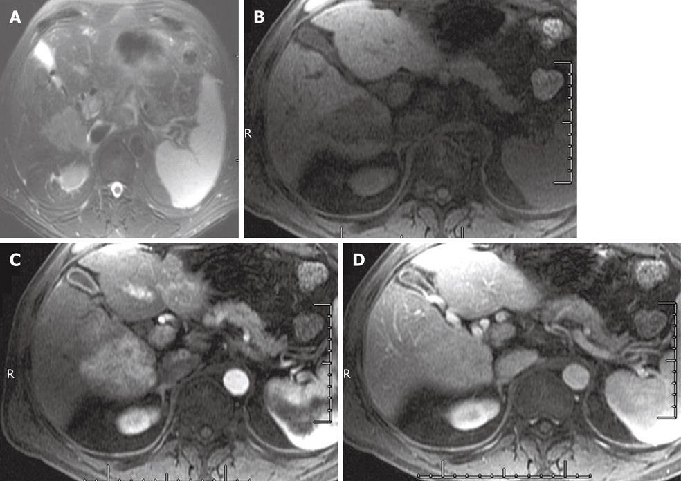
Figure 5 A 65-year-old male with hepatocellular carcinoma.
A: T1 weighted imaging in phase image shows a hypointense mass in the right lobe of the liver; B: T2 weighted imaging (TE = 85 ms) shows a hyperintense mass in the right lobe of the liver; C, D: Late arterial (C) and excretory phase (D) shows a hypervascular mass in the liver that demonstrates an enhancing capsule on the delayed images.
Figure 6 A 62-year-old male with intrahepatic cholangiocarcinoma.
A: Short TE (85 ms) T2 weighted imaging shows a hyperintense mass in the right lobe of the liver; B-D: Pre-contrast (B) late arterial (C), and 5 min delayed images (D) of the abdomen shows delayed enhancement.
Figure 7 A 54-year-old female with intrahepatic cholangiocarcinoma.
The hepatocyte phase of contrast administration shows no uptake of contrast.
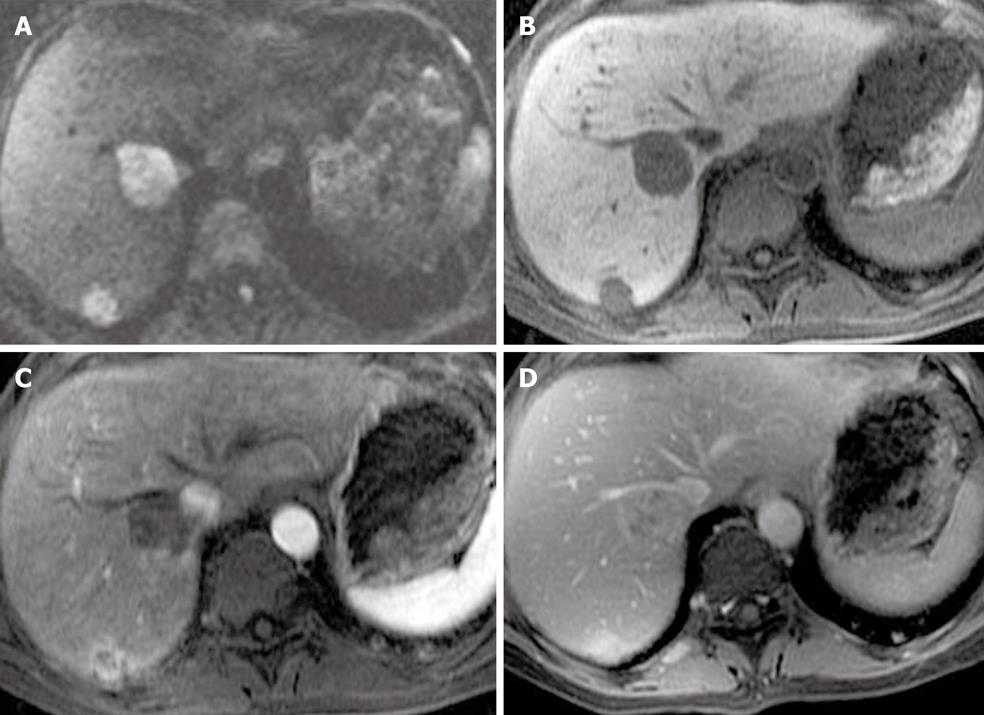
Figure 8 A 51-year-old female with metastatic liver lesions from a pancreatic primary.
A: The diffusion weighted imaging at b = 500 shows two hyperintense nodules in the liver, in keeping with solid lesions; B, C: The late arterial phase (B) shows hypervascular masses that washout on the excretory phase (C) of contrast administration; D: The hepatocyte phase shows low signal lesions in the right lobe of the liver, corresponding to liver metastases.
Figure 9 A 64-year-old with liver metastases from a primary sarcoma.
A: The diffusion weighted imaging at b = 500 shows two solid lesions in the right lobe of the liver; B-D: The pre-contrast (B), late arterial (C), and delayed phase (D) show late enhancement. The enhancement pattern is similar to intrahepatic cholangiocarcinoma.
Figure 10 A 57-year-old female with breast cancer with focal fat deposition in the liver.
The in-phase (A) and out-of-phase (B) images show a signal drop of the area in segment V of the liver.
Figure 11 A 57-year-old female with hepatocellular carcinoma and liver cirrhosis.
The T2 weighted imaging shows enlargement of the lateral segment of the left lobe of the liver and caudate lobe. There is a nodular contour of the liver. All these findings are in keeping with cirrhosis. The spleen is enlarged in keeping with portal hypertension.
Figure 12 A 64-year-old male with a pancreatic mass and iron deposition in the liver.
A: Respiratory triggered fast spin echo T2 weighted imaging image of the liver shows a darker liver relative to the muscles; B, C: In phase (B) and out-of-phase (C) T1 weighted imaging shows a signal drop on the former in keeping with iron deposition.
- Citation: Maniam S, Szklaruk J. Magnetic resonance imaging: Review of imaging techniques and overview of liver imaging. World J Radiol 2010; 2(8): 309-322
- URL: https://www.wjgnet.com/1949-8470/full/v2/i8/309.htm
- DOI: https://dx.doi.org/10.4329/wjr.v2.i8.309


