
















Copyright: ©Author(s) 2026.
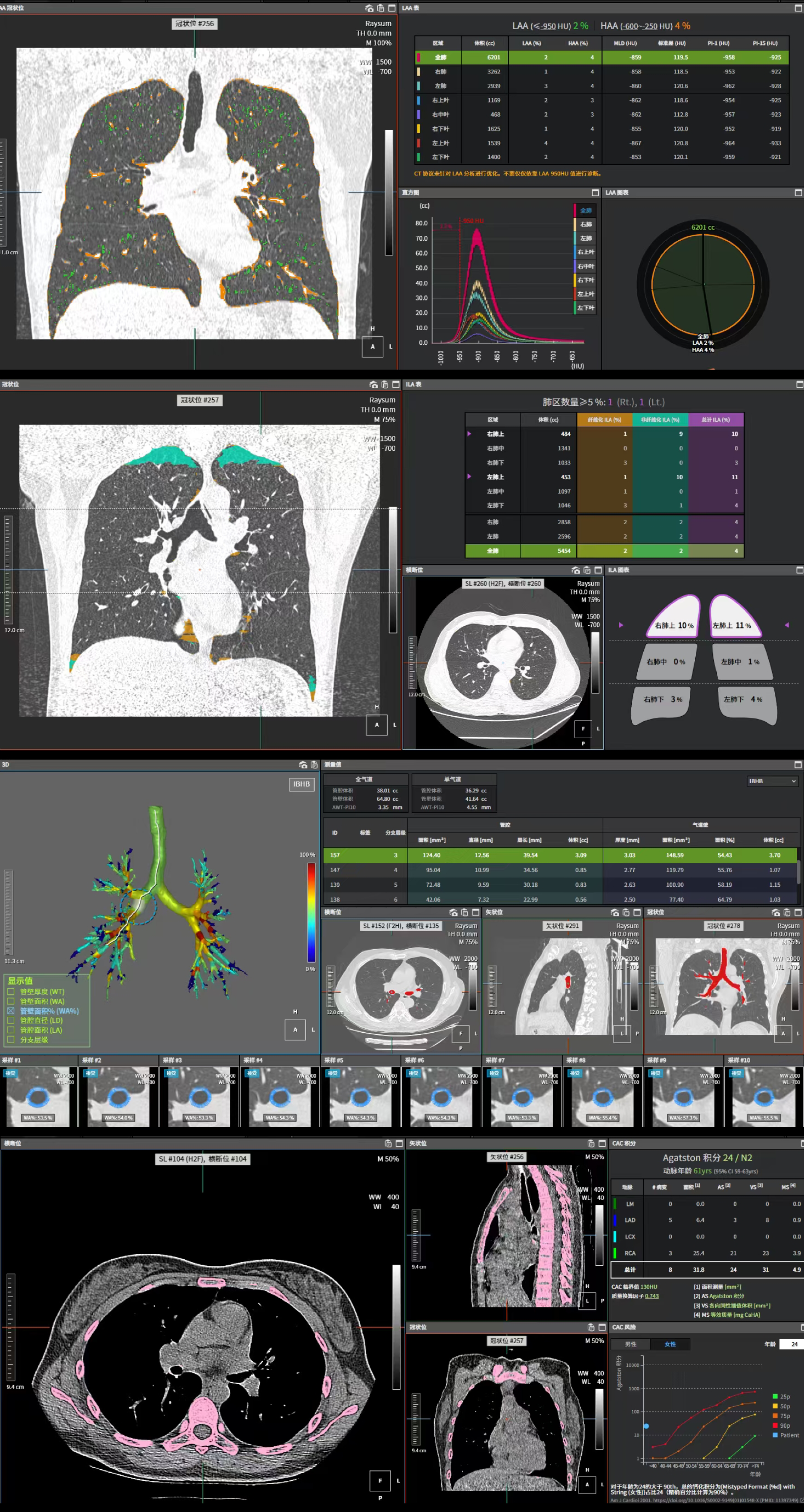
Figure 1 Representative outputs from the SIMBIO-T 1.
0.0 artificial intelligence-based whole-lung quantitative analysis platform. (Hangzhou Shimai Intelligent Technology Co., Ltd.): Low-attenuation area percentage, interstitial lung abnormality, airway wall area percentage, and coronary artery calcification score.
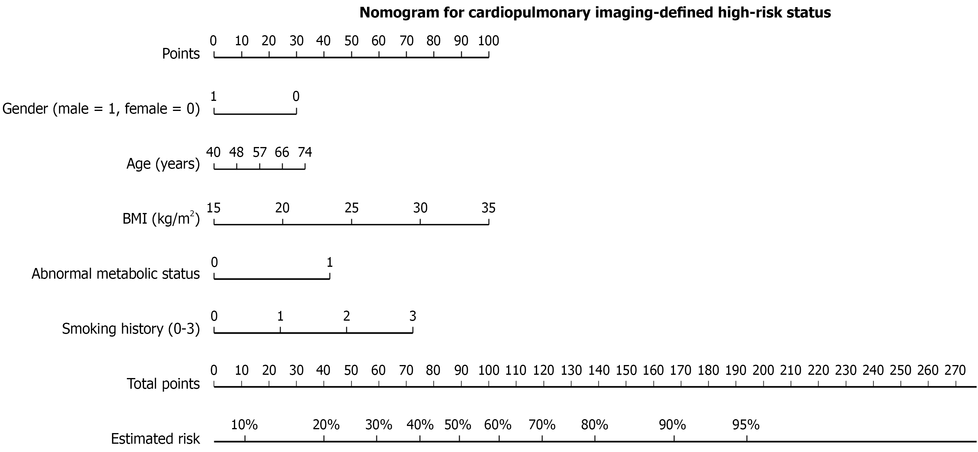
Figure 2 Nomogram of the comprehensive clinical prediction model, including independent risk factors: Sex, age, Body mass index, metabolic status, and smoking history.
Sex (1 = male, 0 = female); metabolic status (1 = abnormal metabolic status, 0 = normal metabolic status); smoking history [0 = non-smoker, 1 = mild (smoking index ≤ 200), 2 = moderate (200 < smoking index < 400), 3 = heavy (smoking index ≥ 400)]. BMI: Body mass index.
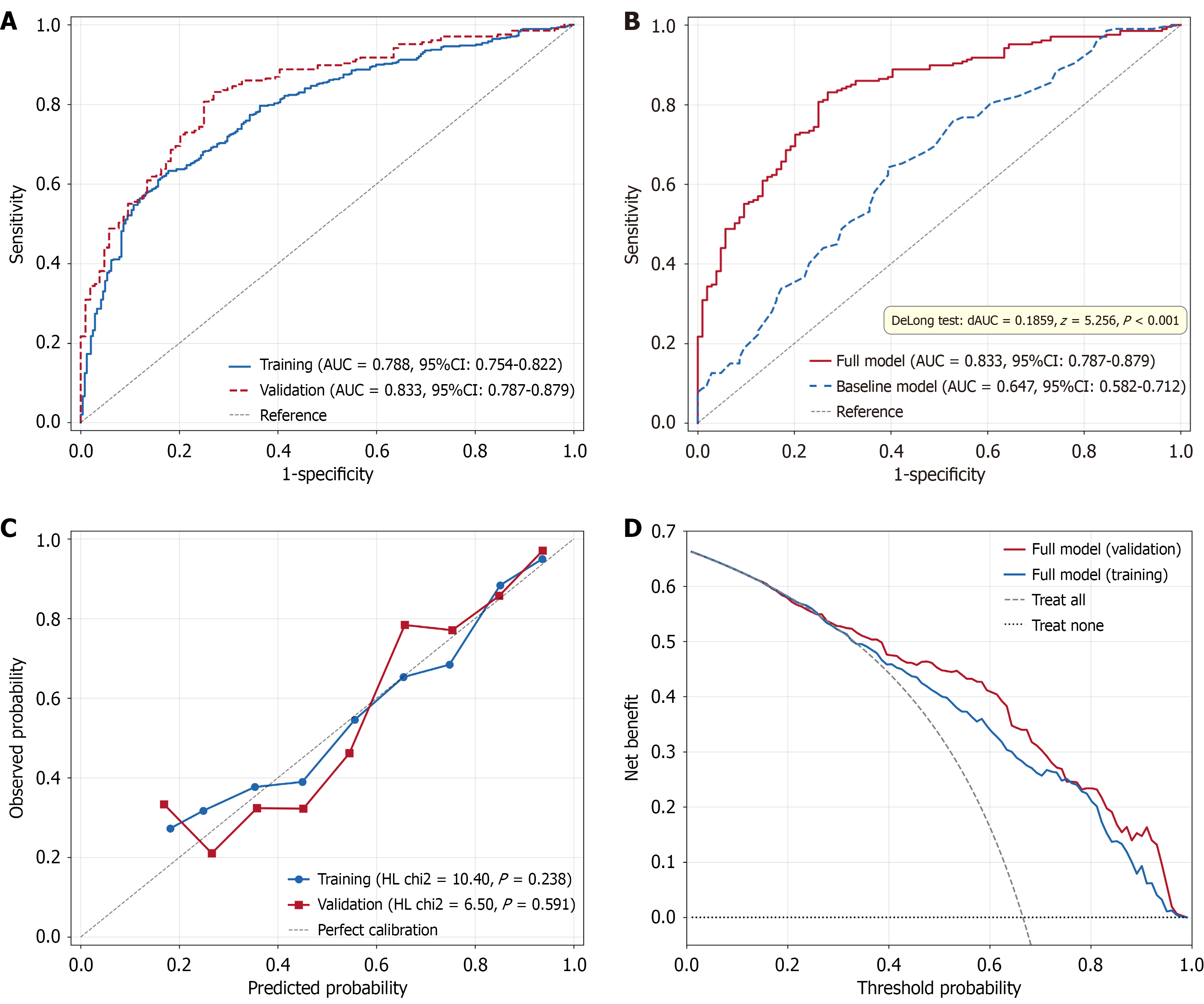
Figure 3 Performance evaluation of the comprehensive clinical prediction model.
A: Receiver operating characteristic curve; B: DeLong test; C: Calibration curves of the training and validation sets; D: Decision curve analysis. AUC: Area under the curve; CI: Confidence interval.
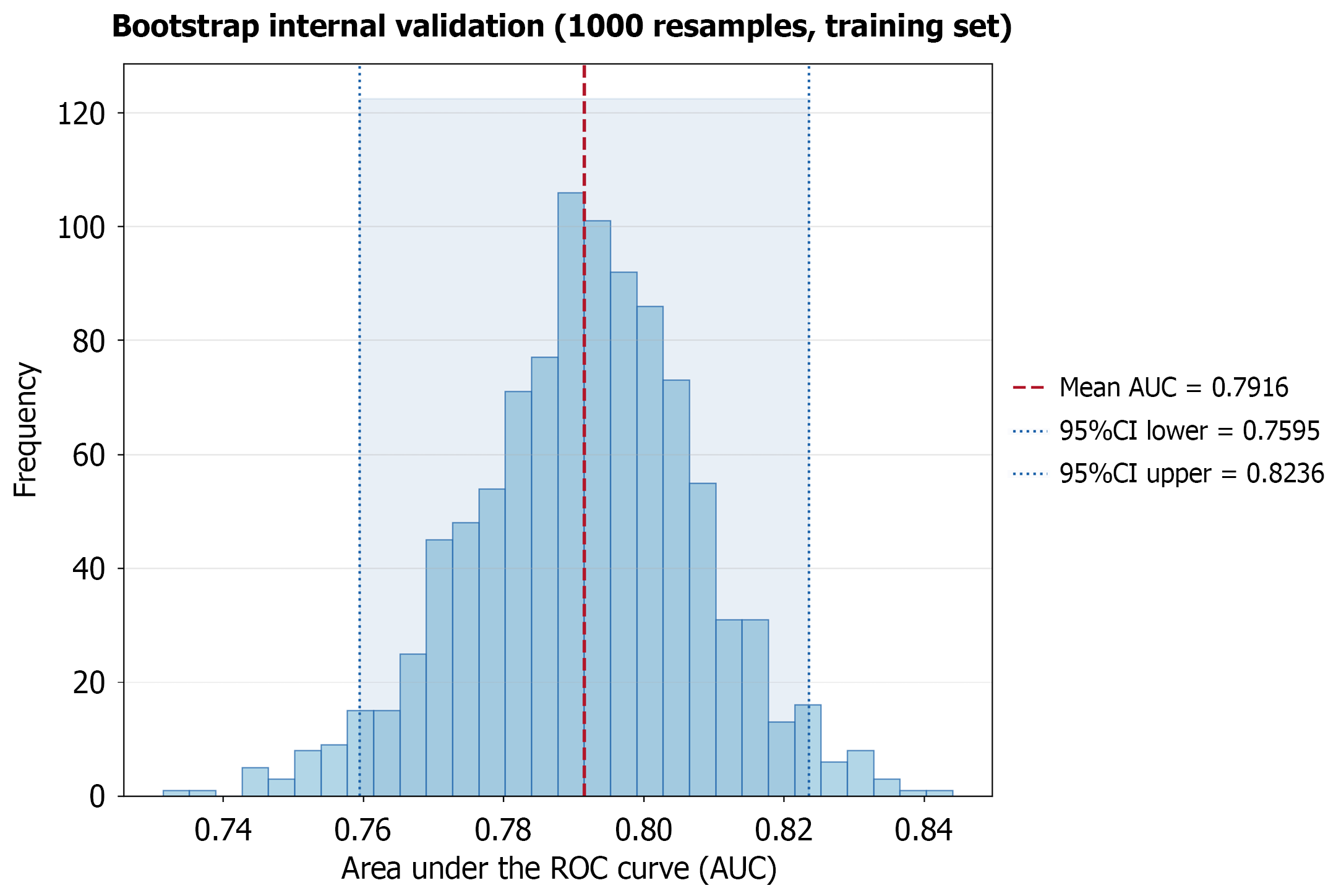
Figure 4 Bootstrap internal resampling validation of the training set model (number of resamplings = 1000).
AUC: Area under the curve; ROC: Receiver operating characteristic.
- Citation: He ZY, Zhan ZW, Zhou LJ, Tang J, Chen C, Yang QJ, Tian Q, Wang XH. Development and validation of a clinical factor-based nomogram for predicting imaging-defined cardiopulmonary abnormality risk in an asymptomatic screening population. World J Radiol 2026; 18(7): 123894
- URL: https://www.wjgnet.com/1949-8470/full/v18/i7/123894.htm
- DOI: https://dx.doi.org/10.4329/wjr.123894