
















Copyright: ©Author(s) 2026.
World J Radiol. May 28, 2026; 18(5): 118851
Published online May 28, 2026. doi: 10.4329/wjr.v18.i5.118851
Published online May 28, 2026. doi: 10.4329/wjr.v18.i5.118851
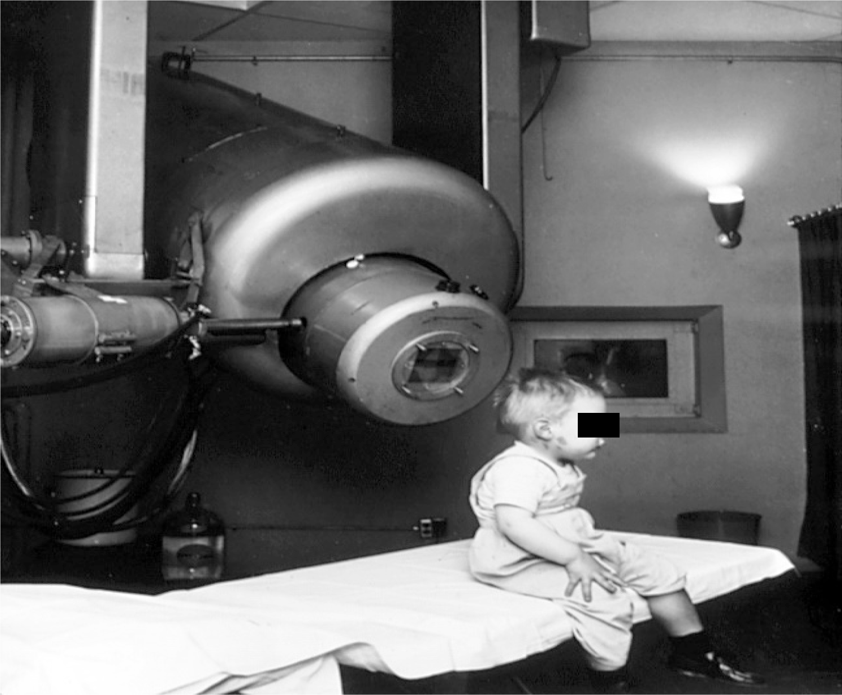
Figure 1 Gordon Isaacs, the first patient treated with the linear accelerator (radiation therapy) for retinoblastoma, sitting on a table[9].
Citation: National Cancer Institute. Linear accelerator to treat retinoblastoma 1957. [cited 5 March 2026]. Available from: https://visualsonline.cancer.gov/details.cfm?imageid=1924. Copyright: ©The Authors 1957. Published by National Cancer Institute Publications. The authors have obtained the permission (Supplementary material).
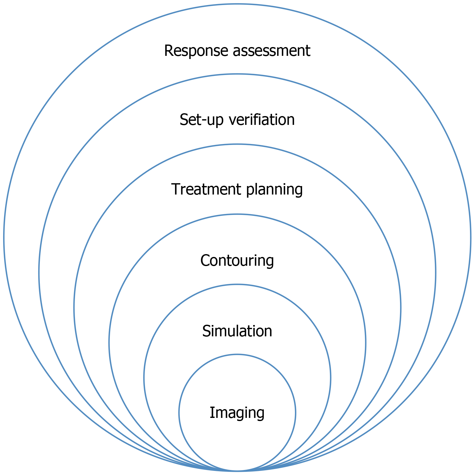
Figure 2
Schematic illustration of the radiotherapy treatment workflow, depicting sequential stages from imaging and simulation to contouring, treatment planning, set-up verification, and treatment response assessment.
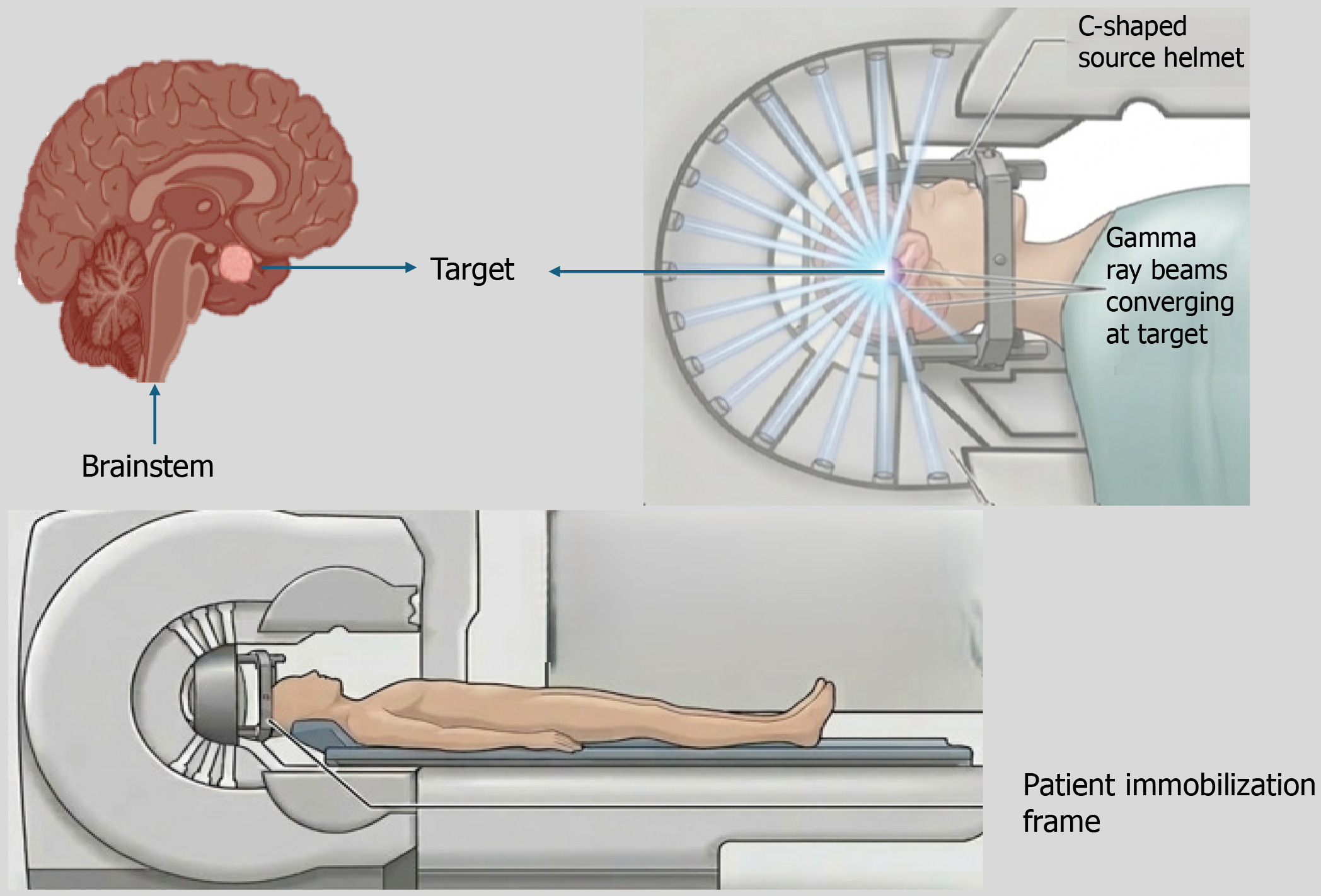
Figure 3
Sectional diagram of gamma knife.
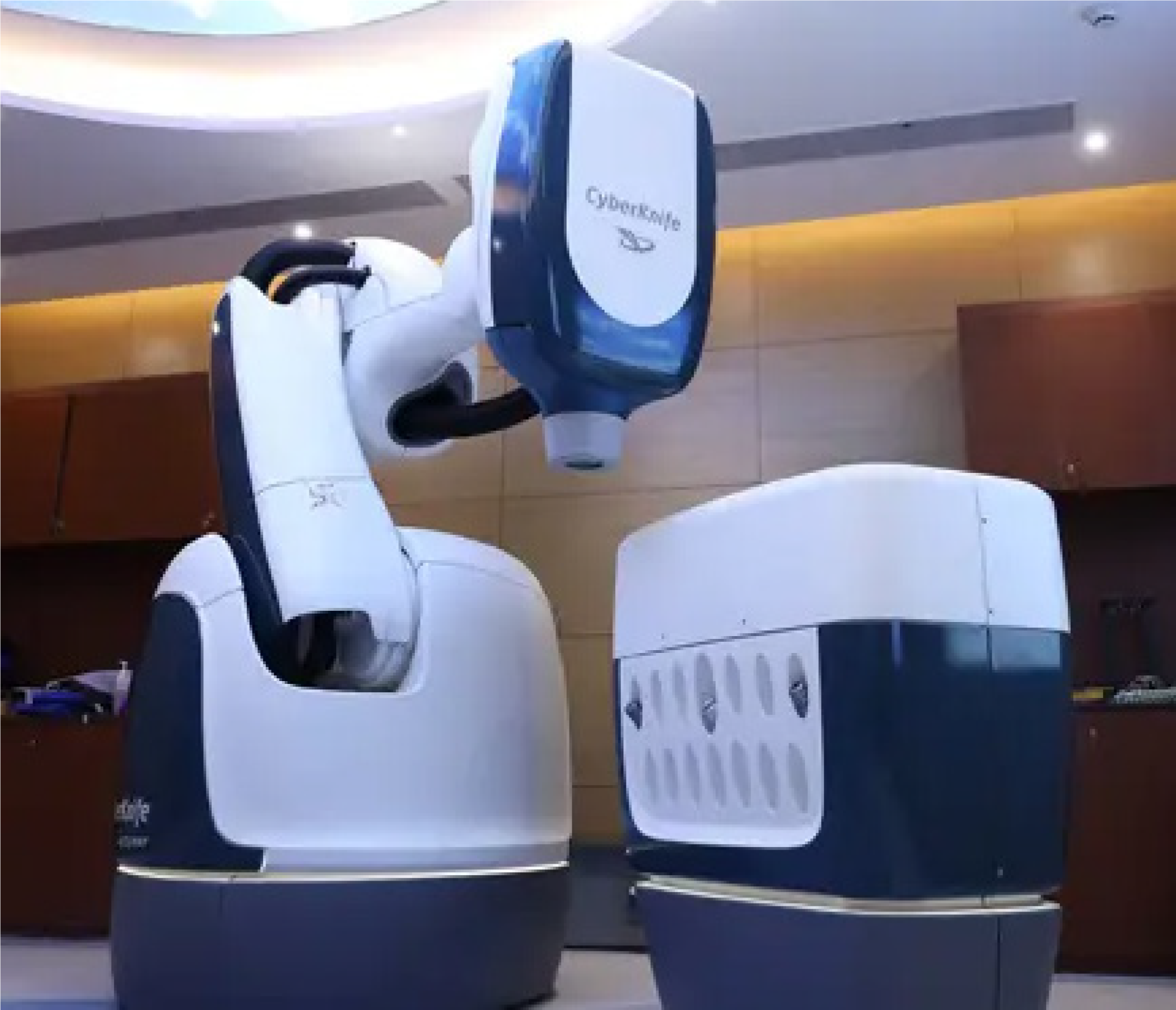
Figure 4
Cyber knife S7 installed at Mahatma Gandhi Medical College and Hospital, Jaipur.
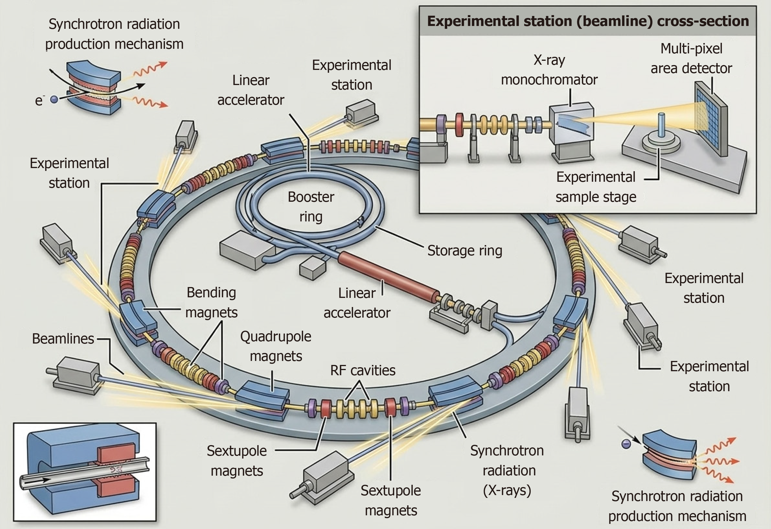
Figure 5
Schematic diagram of synchrotron.
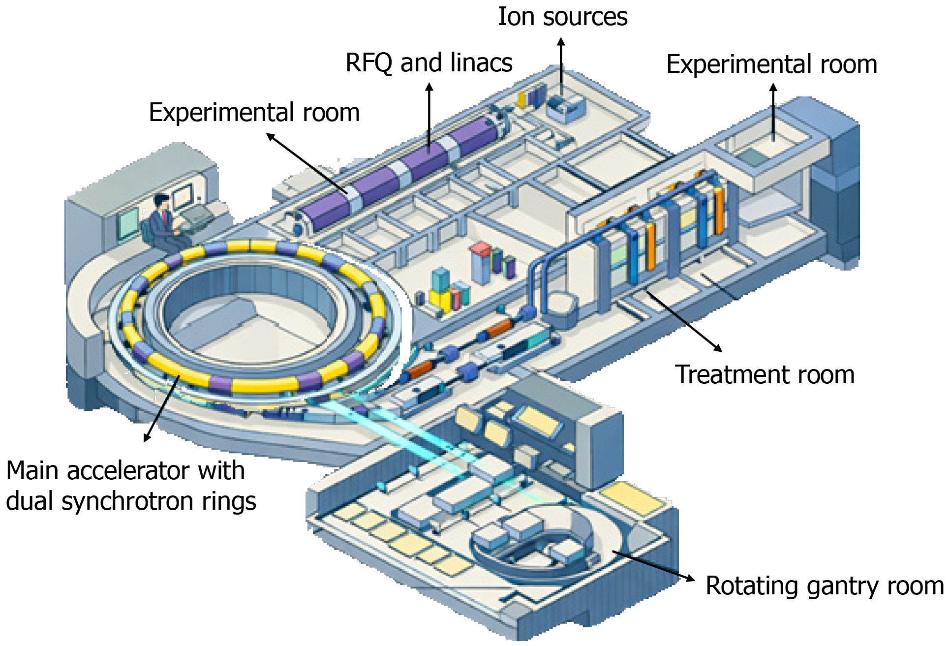
Figure 6 Schematic layout of a heavy ion particle therapy facility.
RFQ: Radio frequency quadruple.
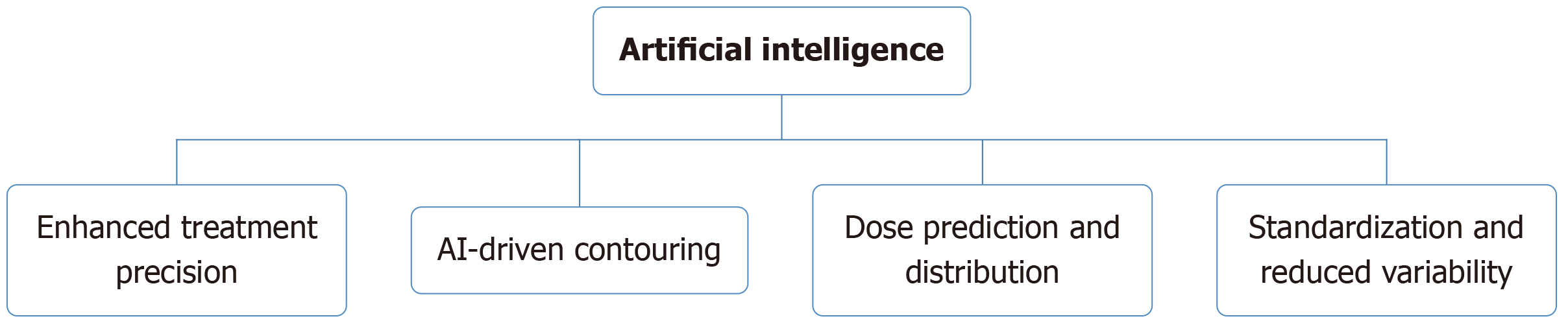
Figure 7
Overview of artificial intelligence applications in radiotherapy, demonstrating its role in automated contouring, dose prediction, treatment precision enhancement, and workflow standardization to reduce clinical variability.
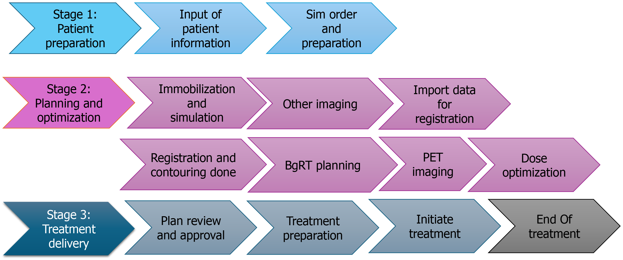
Figure 8 Clinical workflow diagram for biology-guided radiotherapy.
PET: Positron emission tomography; BGRT: Biologically guided radiation therapy; EOT: End-of-treatment.
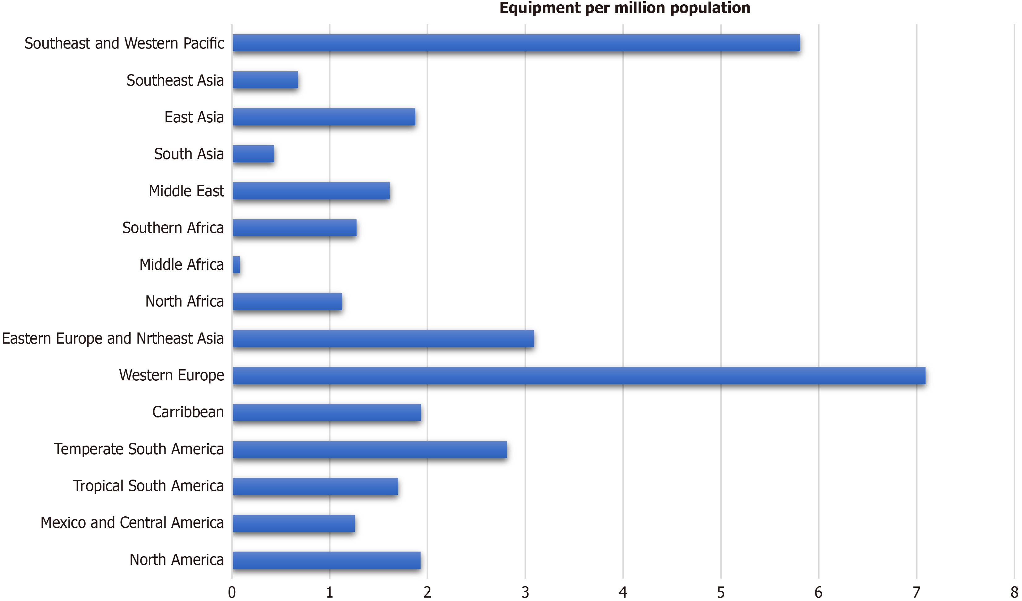
Figure 9
Regional distribution of radiotherapy equipment per million population worldwide, highlighting significant disparities in radiotherapy infrastructure availability between high-income and low- and middle-income regions.
- Citation: Singh P, Singh MK, Mishra A. Journey through technological advancements in radiation therapy. World J Radiol 2026; 18(5): 118851
- URL: https://www.wjgnet.com/1949-8470/full/v18/i5/118851.htm
- DOI: https://dx.doi.org/10.4329/wjr.v18.i5.118851