
















Copyright: ©Author(s) 2026.
World J Radiol. May 28, 2026; 18(5): 117936
Published online May 28, 2026. doi: 10.4329/wjr.v18.i5.117936
Published online May 28, 2026. doi: 10.4329/wjr.v18.i5.117936
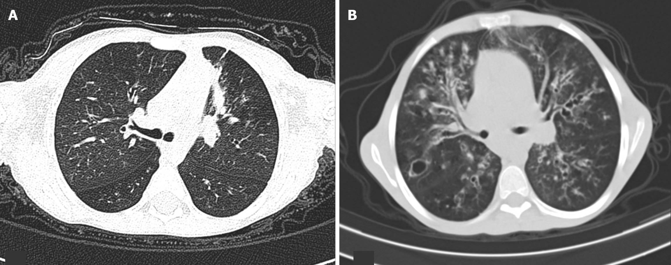
Figure 1 Depicting progressive deterioration of the lungs on high-resolution computed tomography.
A: Baseline high-resolution computed tomography (HRCT) shows bronchial wall thickening with subsegmental collapse of the anterior segment of the left upper lobe, along with small centrilobular nodules; B: Follow-up HRCT after one year demonstrates extensive cylindrical, varicoid, and cystic bronchiectasis involving both lungs, with associated bronchial wall thickening and multiple air-space nodules.
- Citation: Suhail JM, Wagay MI, Parry AH, Bhat IM, Sheikh RR, Bhat MA. Radiologic progression with minimal functional decline in cystic fibrosis: Insights from a prospective study. World J Radiol 2026; 18(5): 117936
- URL: https://www.wjgnet.com/1949-8470/full/v18/i5/117936.htm
- DOI: https://dx.doi.org/10.4329/wjr.v18.i5.117936