
















Copyright: ©Author(s) 2026.
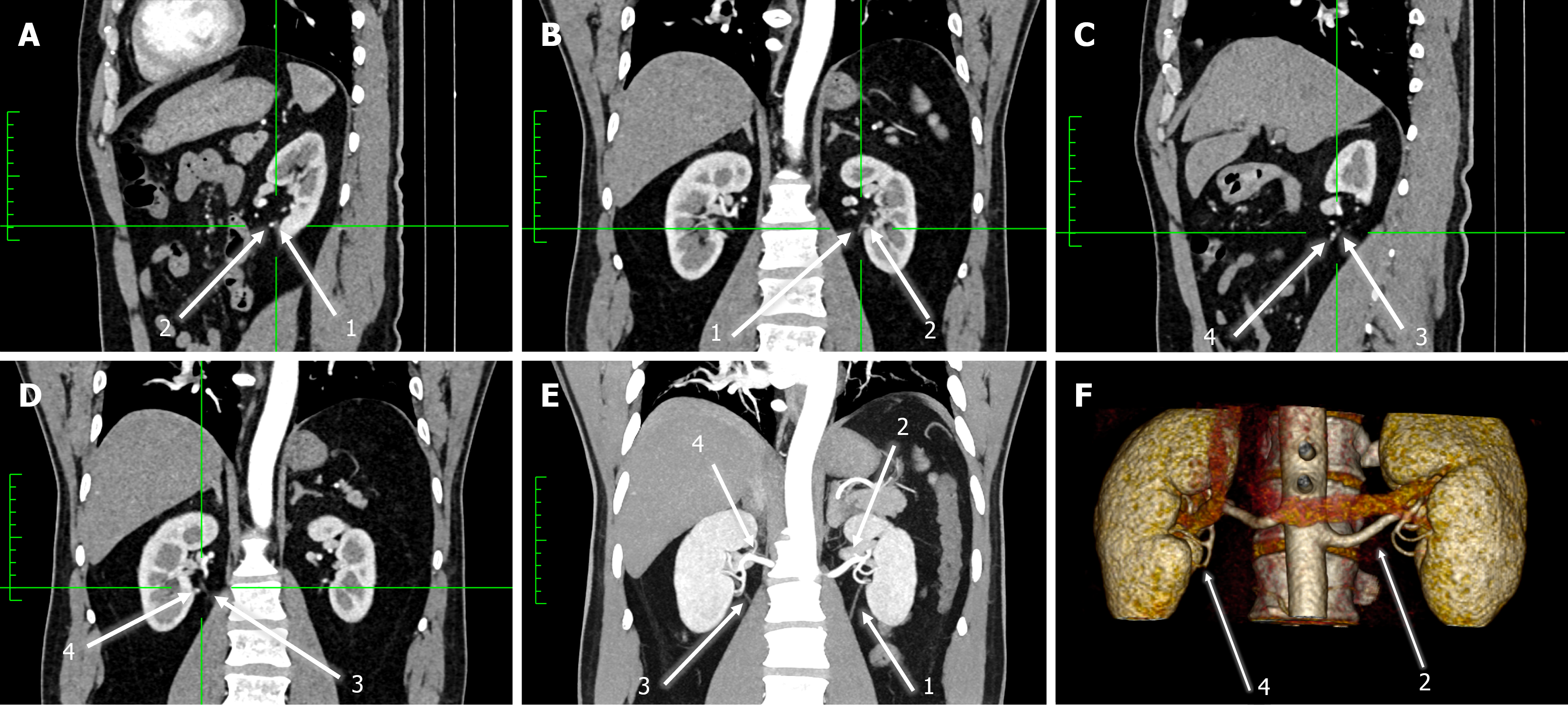
Figure 1 Computed tomography scan of 74-year-old female patient exhibiting anterior crossing by both early bifurcated renal artery and vein at the level of the uteropelvic junction.
A, B: Left-sided; C, D: Right-sided; E: Bilateral maximum-intensity projection; F: Three-dimensional reconstructions. A, C: Sagittal; B, D, and E: Coronal. 1: Left Ureter; 2: Left early bifurcated renal arteries; 3: Right Ureter; 4: Right Early Bifurcated Renal artery.
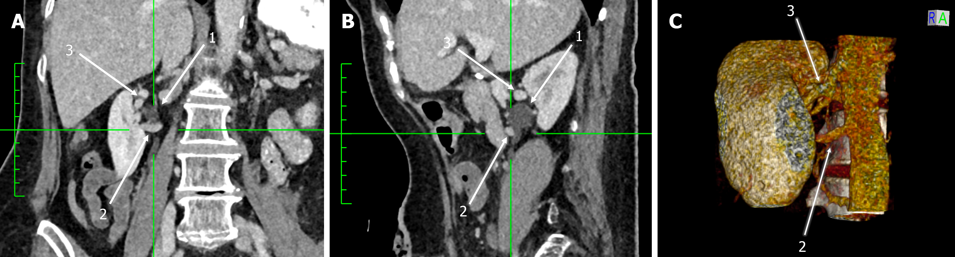
Figure 2 Computed tomography scan of 54-year-old male patient exhibiting bilateral anterior crossing by early bifurcated renal arteries at the level of ureteropelvic junction (on the left side) and 12.
5 mm distally on the ureter (on the right side). A: Coronal; B: Sagittal; C: Three-dimensional reconstruction. 1: Left ureter, 2: Left early bifurcated renal artery; 3: Right ureter.
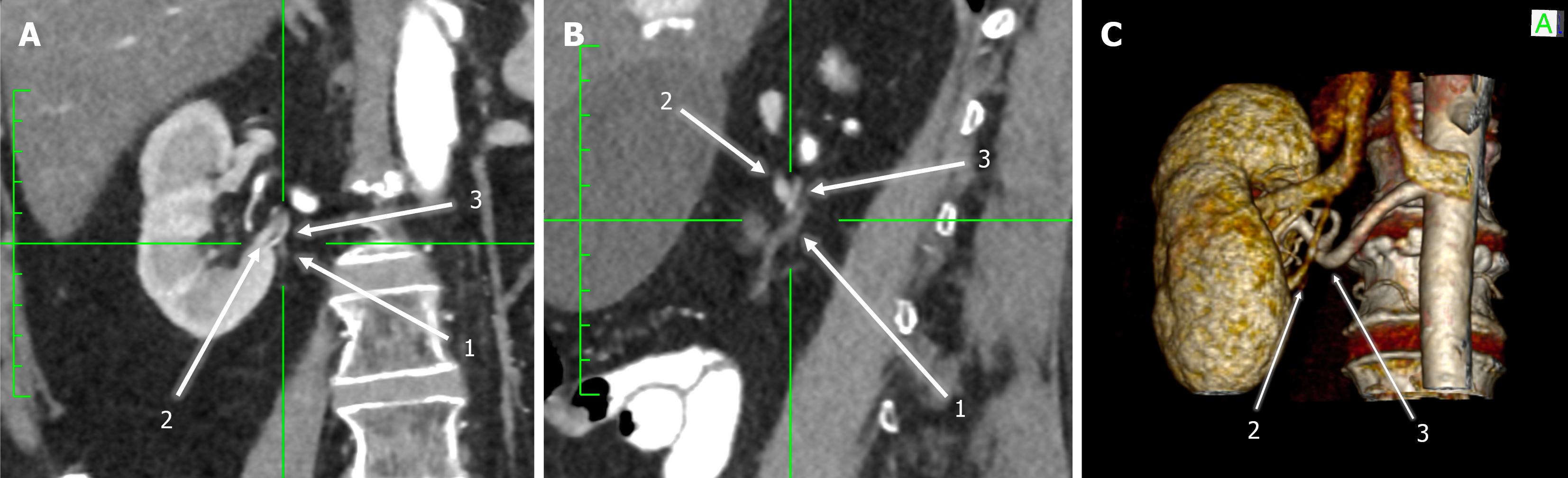
Figure 3 Computed tomography scan of 55-year-old male patient exhibiting anterior crossing by an accessory renal artery, 19.
9 mm distally to the ureter. A: Coronal; B: Sagittal; C: Three-dimensional reconstructions. 1: Ureter; 2: Typical renal artery; 3: Accessory renal artery.
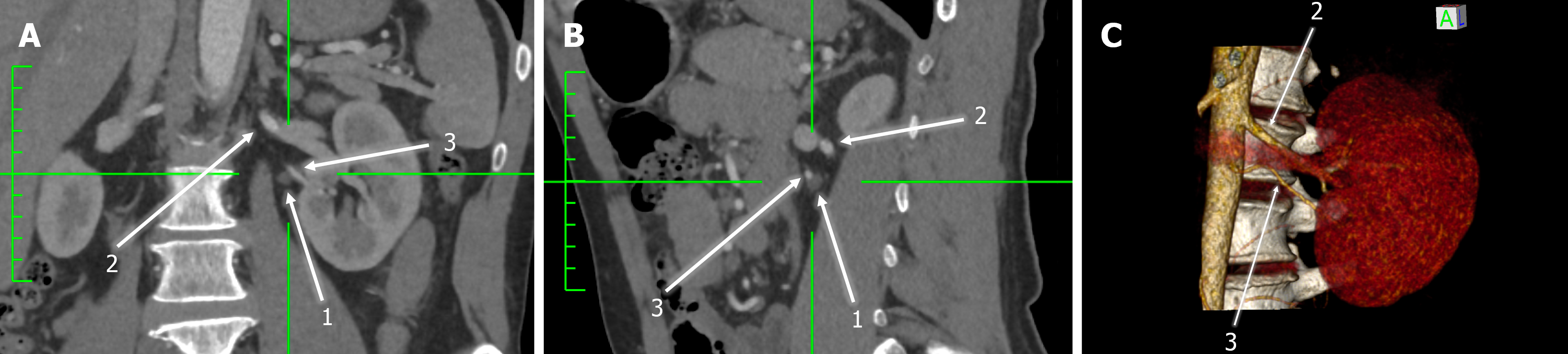
Figure 4 Computed tomography scan of 64-year-old female patient exhibiting anterior crossing by an accessory renal vein at the level of ureteropelvic junction.
A: Coronal; B: Sagittal; C: Three-dimensional reconstruction. 1: Ureter; 2: Accessory renal vein; 3: Typical renal vein.
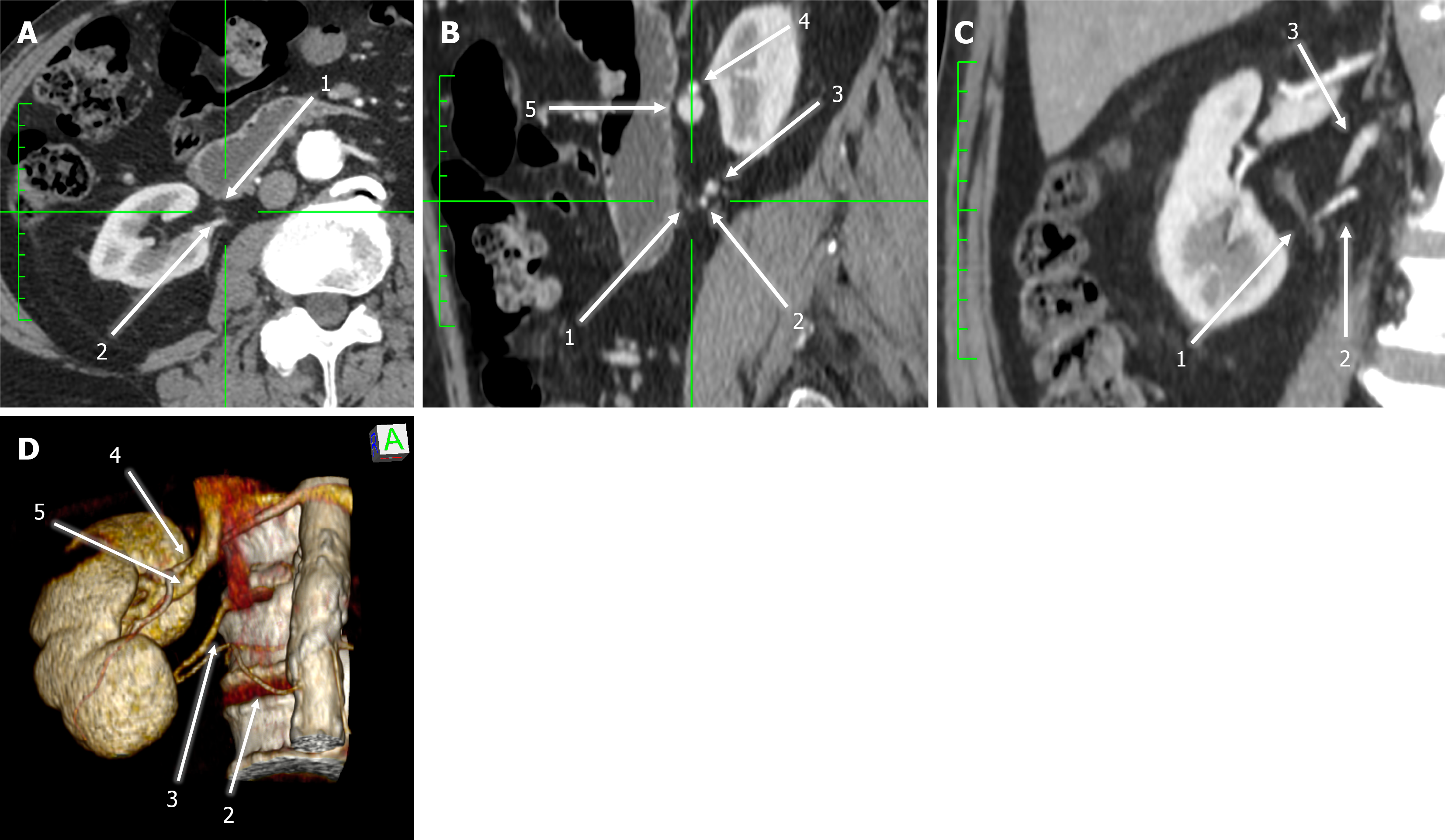
Figure 5 Computed tomography scan of 65-year-old male patient exhibiting posterior crossing by both accessory renal artery and vein, 15.
1 mm distally to the ureter. A: Axial; B: Sagittal; C: Coronal; D: Three-dimensional reconstructions. 1: Ureter; 2: Accessory renal artery; 3: Accessory renal vein; 4: Typical renal artery; 5: Typical renal vein.
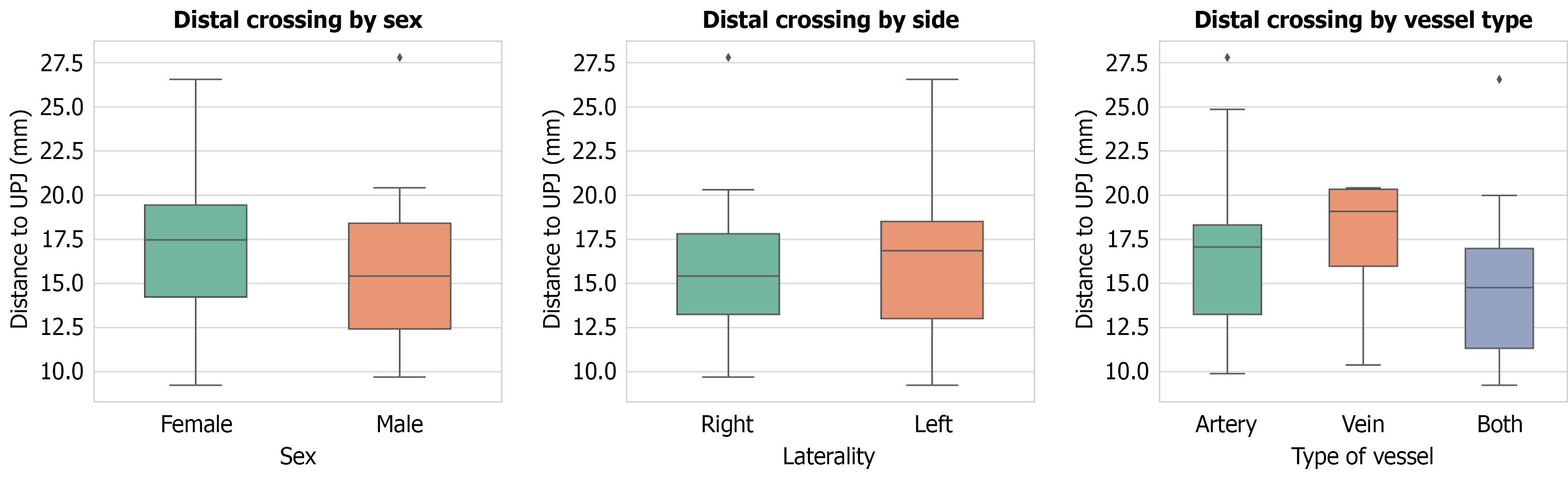
Figure 6 Box plots demonstrating the distal site of crossing (> 0 mm) between the vessel and the ureteropelvic junction, categorized by patient sex.
Left: Female (n = 17), male (n = 39), laterality [center: Left (n = 31), right (n = 25)], and vascular etiology [right: Artery (n = 36), vein (n = 6), both (n = 14)]. The central horizontal line within each box represents the median distance, while the lower and upper boundaries of the box indicate the first and third quartiles (interquartile range). The whiskers extend to the lowest and highest values within 1.5 times the interquartile range. Diamond markers denote outlier data points beyond this range.
- Citation: Paschopoulos I, Triantafyllou G, Gondorf D, Arkoudis NA, Velonakis G, Mariorakis C, Lyrtzis C, Gyftopoulos K, Piagkou M. Aberrant renal vessels crossing the ureter and the ureteropelvic junction: A retrospective computed tomography-based analysis of non-obstructed population. World J Radiol 2026; 18(10): 127348
- URL: https://www.wjgnet.com/1949-8470/full/v18/i10/127348.htm
- DOI: https://dx.doi.org/10.4329/wjr.127348