
















Copyright: ©Author(s) 2026.
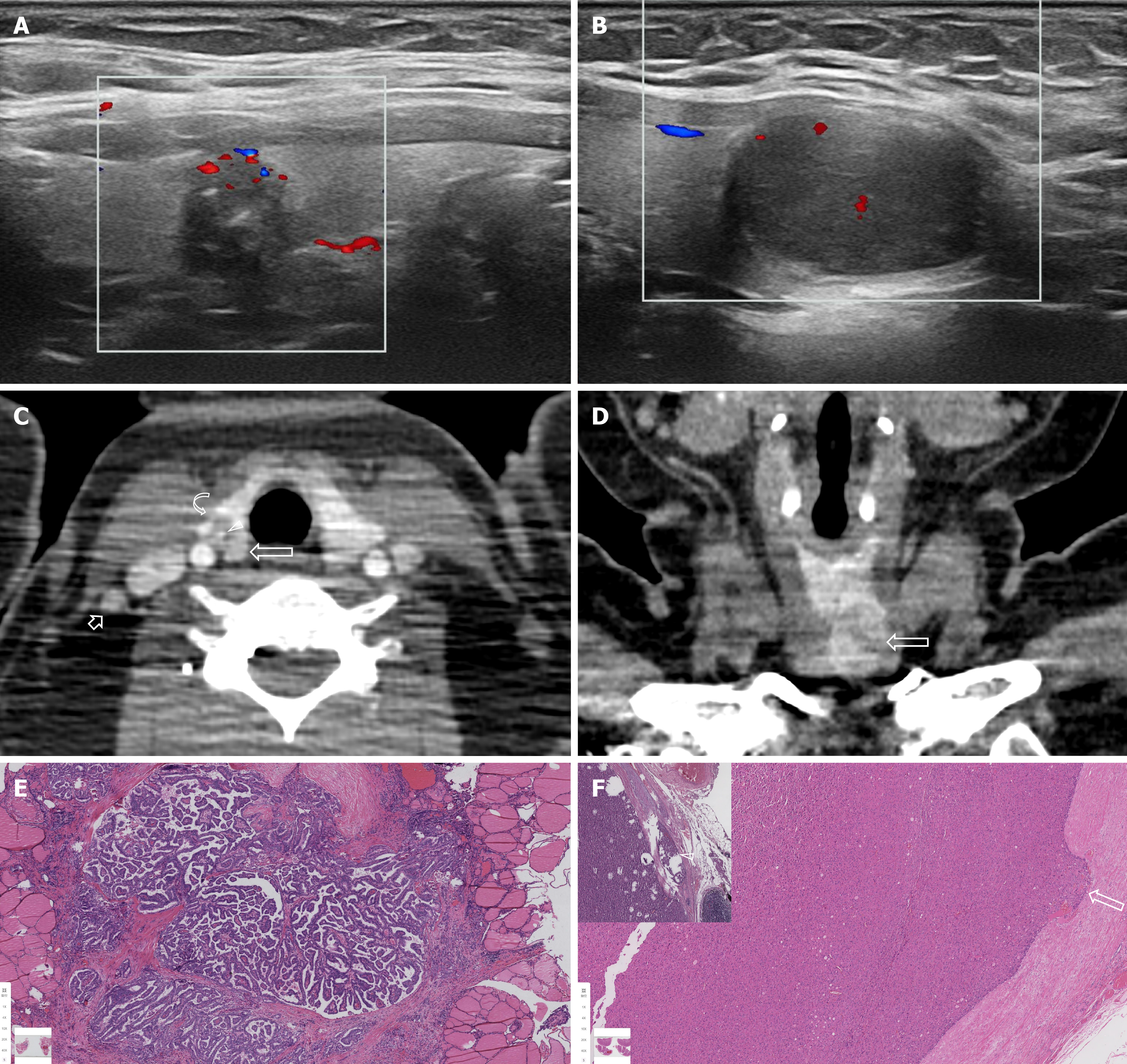
Figure 1 A 38-year-old female with collision tumors comprising oncocytic and papillary thyroid carcinomas.
A: Ultrasound showed an irregular nodule with abundant intravascular in the right thyroid lobe [Chinese Thyroid Imaging Reporting and Data System (C-TIRADS) 4A]; B: Ultrasound showed a well-demarcated irregular nodule with some intravascular in the left thyroid lobe (C-TIRADS 3); C: Contrast-enhanced computed tomography (CT) revealed an ill-demarcated node (long arrow) with heterogeneous enhancement, calcification (arrowhead) and suspected minimal extrathyroidal extension (curve arrow) in the right thyroid lobe. A lymph node (short arrow) with avid enhancement was noted in the right lateral compartment; D: Contrast-enhanced coronal CT showed a well-demarcated node with nonuniform enhancement in the left thyroid lobe (arrow); E: Histologic section [hematoxylin and eosin (H&E) staining, × 5 original magnification] of the right lobe node indicated classical papillary thyroid carcinoma with encapsulated invasion and no vascular invasion; F: Histologic section (H&E staining, × 5 original magnification) of the left lobe node indicated tumor cells with avid oncocytic staining, encapsulated invasion (arrow), and vascular invasion (arrowhead in the inset).
- Citation: Gong QS, Liu XW, Luo XP, Xu N. Imaging features of synchronous bilateral primary tumors of oncocytic thyroid carcinoma and papillary thyroid carcinoma: A case report. World J Radiol 2026; 18(10): 126932
- URL: https://www.wjgnet.com/1949-8470/full/v18/i10/126932.htm
- DOI: https://dx.doi.org/10.4329/wjr.126932